

with up to 40 % mortality, although other organs may also be
affected. Affected fish show unspecific clinical signs that
may include cataract, snout ulcers and increased numbers
of ‘poor doing fish’. At autopsy, the pseudobranch is
haemorrhagic or totally absent, leaving only a white
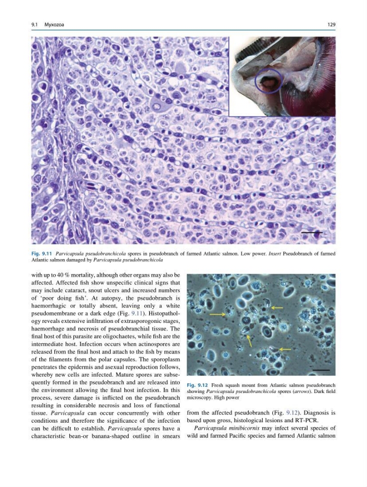
pseudomembrane or a dark edge (Fig. 9.11). Histopathology reveals extensive infiltration of extrasporogonic stages,
haemorrhage and necrosis of pseudobranchial tissue. The
final host of this parasite are oligochaetes, while fish are the
intermediate host. Infection occurs when actinospores are
released from the final host and attach to the fish by means
of the filaments from the polar capsules. The sporoplasm
penetrates the epidermis and asexual reproduction follows,
whereby new cells are infected. Mature spores are subsequently formed in the pseudobranch and are released into
the environment allowing the final host infection. In this
process, severe damage is inflicted on the pseudobranch
resulting in considerable necrosis and loss of functional
tissue. Parvicapsula can occur concurrently with other
conditions and therefore the significance of the infection
can be difficult to establish. Parvicapsula spores have a
characteristic bean-or banana-shaped outline in smears
from the affected pseudobranch (Fig. 9.12). Diagnosis is
based upon gross, histological lesions and RT-PCR.
Parvicapsula minibicornis may infect several species of
wild and farmed Pacific species and farmed Atlantic salmon
Fig. 9.11 Parvicapsula pseudobranchicola spores in pseudobranch of farmed Atlantic salmon. Low power. Insert Pseudobranch of farmed
Atlantic salmon damaged by Parvicapsula pseudobranchicola
Fig. 9.12 Fresh squash mount from Atlantic salmon pseudobranch
showing Parvicapsula pseudobranchicola spores (arrows). Dark field
microscopy. High power
9.1 Myxozoa
129
affected. Affected fish show unspecific clinical signs that
may include cataract, snout ulcers and increased numbers
of ‘poor doing fish’. At autopsy, the pseudobranch is
haemorrhagic or totally absent, leaving only a white
pseudomembrane or a dark edge (Fig. 9.11). Histopathology reveals extensive infiltration of extrasporogonic stages,
haemorrhage and necrosis of pseudobranchial tissue. The
final host of this parasite are oligochaetes, while fish are the
intermediate host. Infection occurs when actinospores are
released from the final host and attach to the fish by means
of the filaments from the polar capsules. The sporoplasm
penetrates the epidermis and asexual reproduction follows,
whereby new cells are infected. Mature spores are subsequently formed in the pseudobranch and are released into
the environment allowing the final host infection. In this
process, severe damage is inflicted on the pseudobranch
resulting in considerable necrosis and loss of functional
tissue. Parvicapsula can occur concurrently with other
conditions and therefore the significance of the infection
can be difficult to establish. Parvicapsula spores have a
characteristic bean-or banana-shaped outline in smears
from the affected pseudobranch (Fig. 9.12). Diagnosis is
based upon gross, histological lesions and RT-PCR.
Parvicapsula minibicornis may infect several species of
wild and farmed Pacific species and farmed Atlantic salmon
Fig. 9.11 Parvicapsula pseudobranchicola spores in pseudobranch of farmed Atlantic salmon. Low power. Insert Pseudobranch of farmed
Atlantic salmon damaged by Parvicapsula pseudobranchicola
Fig. 9.12 Fresh squash mount from Atlantic salmon pseudobranch
showing Parvicapsula pseudobranchicola spores (arrows). Dark field
microscopy. High power
9.1 Myxozoa
129