

Spores are released from the cartilage after death of
the fish where they are ingested by an intermediate host,
the oligochaete Tubifex tubiflex, in which they localize in the
intestinal epithelium and develop into the triactinomyxon
stage. Re-infection in the fish host may occur through the
skin and buccal cavity. Trophozoites migrate through the
epidermis or gut lining and peripheral nerves to reach
the cranial cartilage. In rainbow trout successful development of the parasite only occurs in sac fry which are older
than 2 days. Trophozoites induce necrosis either directly or
indirectly and lysis in the cartilage of the head and vertebrae.
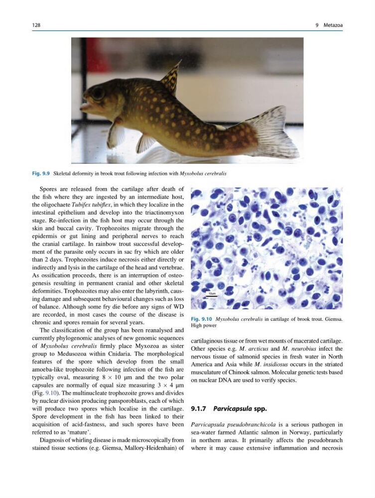
As ossification proceeds, there is an interruption of osteogenesis resulting in permanent cranial and other skeletal
deformities. Trophozoites may also enter the labyrinth, causing damage and subsequent behavioural changes such as loss
of balance. Although some fry die before any signs of WD
are recorded, in most cases the course of the disease is
chronic and spores remain for several years.
The classification of the group has been reanalysed and
currently phylogenomic analyses of new genomic sequences
of Myxobolus cerebralis firmly place Myxozoa as sister
group to Medusozoa within Cnidaria. The morphological
features of the spore which develop from the small
amoeba-like trophozoite following infection of the fish are
typically oval, measuring 8 Â 10 μm and the two polar
capsules are normally of equal size measuring 3 Â 4 μm
(Fig. 9.10). The multinucleate trophozoite grows and divides
by nuclear division producing pansporoblasts, each of which
will produce two spores which localise in the cartilage.
Spore development in the fish has been linked to their
acquisition of acid-fastness, and such spores have been
referred to as ‘mature’.
Diagnosis of whirling disease is made microscopically from
stained tissue sections (e.g. Giemsa, Mallory-Heidenhain) of
cartilaginous tissue or from wet mounts of macerated cartilage.
Other species e.g. M. arcticus and M. neurobius infect the
nervous tissue of salmonid species in fresh water in North
America and Asia while M. insidiosus occurs in the striated
musculature of Chinook salmon. Molecular genetic tests based
on nuclear DNA are used to verify species.
9.1.7 Parvicapsula spp.
Parvicapsula pseudobranchicola is a serious pathogen in
sea-water farmed Atlantic salmon in Norway, particularly
in northern areas. It primarily affects the pseudobranch
where it may cause extensive inflammation and necrosis
Fig. 9.9 Skeletal deformity in brook trout following infection with Myxobolus cerebralis
Fig. 9.10 Myxobolus cerebralis in cartilage of brook trout. Giemsa.
High power
128
9 Metazoa
the fish where they are ingested by an intermediate host,
the oligochaete Tubifex tubiflex, in which they localize in the
intestinal epithelium and develop into the triactinomyxon
stage. Re-infection in the fish host may occur through the
skin and buccal cavity. Trophozoites migrate through the
epidermis or gut lining and peripheral nerves to reach
the cranial cartilage. In rainbow trout successful development of the parasite only occurs in sac fry which are older
than 2 days. Trophozoites induce necrosis either directly or
indirectly and lysis in the cartilage of the head and vertebrae.
As ossification proceeds, there is an interruption of osteogenesis resulting in permanent cranial and other skeletal
deformities. Trophozoites may also enter the labyrinth, causing damage and subsequent behavioural changes such as loss
of balance. Although some fry die before any signs of WD
are recorded, in most cases the course of the disease is
chronic and spores remain for several years.
The classification of the group has been reanalysed and
currently phylogenomic analyses of new genomic sequences
of Myxobolus cerebralis firmly place Myxozoa as sister
group to Medusozoa within Cnidaria. The morphological
features of the spore which develop from the small
amoeba-like trophozoite following infection of the fish are
typically oval, measuring 8 Â 10 μm and the two polar
capsules are normally of equal size measuring 3 Â 4 μm
(Fig. 9.10). The multinucleate trophozoite grows and divides
by nuclear division producing pansporoblasts, each of which
will produce two spores which localise in the cartilage.
Spore development in the fish has been linked to their
acquisition of acid-fastness, and such spores have been
referred to as ‘mature’.
Diagnosis of whirling disease is made microscopically from
stained tissue sections (e.g. Giemsa, Mallory-Heidenhain) of
cartilaginous tissue or from wet mounts of macerated cartilage.
Other species e.g. M. arcticus and M. neurobius infect the
nervous tissue of salmonid species in fresh water in North
America and Asia while M. insidiosus occurs in the striated
musculature of Chinook salmon. Molecular genetic tests based
on nuclear DNA are used to verify species.
9.1.7 Parvicapsula spp.
Parvicapsula pseudobranchicola is a serious pathogen in
sea-water farmed Atlantic salmon in Norway, particularly
in northern areas. It primarily affects the pseudobranch
where it may cause extensive inflammation and necrosis
Fig. 9.9 Skeletal deformity in brook trout following infection with Myxobolus cerebralis
Fig. 9.10 Myxobolus cerebralis in cartilage of brook trout. Giemsa.
High power
128
9 Metazoa