

During the life cycle, actinospores are discharged from an
annelid host into the water and penetrate the skin of the fish
intermediate host. The sporoplasm develops into presporogonic stages and eventually will locate in the bile
ducts where the sporogonic stage, plasmodium develops.
The cytoplasm of these large worm- or sac-like structures
is filled with many cell types including sporogonic cells and
pericytes. These develop into sporogony (spore formation),
and subsequently into sporoblasts and myxospores.
Histological examination reveals dilated bile ducts and
excretory canals with characteristic worm-like plasmodia
filled with sporogonic cells and spores (Fig. 9.7). Diagnosis
is based upon the examination of wet mounts or tissue sections,
the identification of typical plasmodia and crescent shaped or
fusiform spores and polar capsules at opposing ends.
9.1.6 Myxobolus cerebralis
Myxobolus cerebralis is the causal agent of a persistent and
economical important condition termed ‘whirling disease’
(WD). The parasite has been identified from farmed and wild
Pacific and Atlantic salmon, but within farmed species this is
mainly a problem in trout reared in earthen ponds. Clinical
signs include spiralling (‘whirling behaviour’), darkening of
the caudal region and severe skeletal deformities of the
cranial area, jaw and opercula (Figs. 9.8 and 9.9) The
whirling behaviour is often pronounced when the fish
attempt to feed or when the fish are startled. Erosion of the
cartilage surrounding the auditory organ is reported to contribute to the whirling. Infection of the cartilage of the spinal
column causes pressure on the caudal nerves resulting in loss
of control of caudal dermal melanophores. Infections cause a
significant pathology usually in the first 3–4 months after
fish start feeding and before ossification is complete. Maturing stages lyse and digest chondrocytes thus disrupting osteogenesis. Thereafter, older fish may be infected but
infections are unlikely to result in a clinical complication.
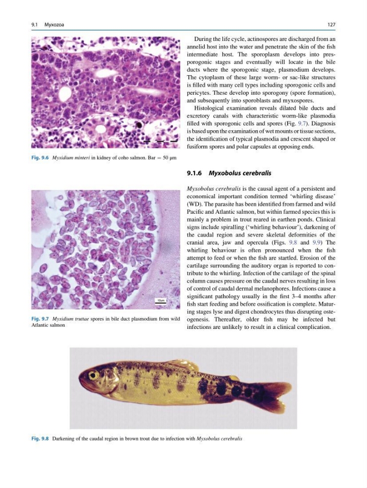
Fig. 9.6 Myxidium minteri in kidney of coho salmon. Bar ¼ 50 μm
Fig. 9.7 Myxidium truttae spores in bile duct plasmodium from wild
Atlantic salmon
Fig. 9.8 Darkening of the caudal region in brown trout due to infection with Myxobolus cerebralis
9.1 Myxozoa
127
annelid host into the water and penetrate the skin of the fish
intermediate host. The sporoplasm develops into presporogonic stages and eventually will locate in the bile
ducts where the sporogonic stage, plasmodium develops.
The cytoplasm of these large worm- or sac-like structures
is filled with many cell types including sporogonic cells and
pericytes. These develop into sporogony (spore formation),
and subsequently into sporoblasts and myxospores.
Histological examination reveals dilated bile ducts and
excretory canals with characteristic worm-like plasmodia
filled with sporogonic cells and spores (Fig. 9.7). Diagnosis
is based upon the examination of wet mounts or tissue sections,
the identification of typical plasmodia and crescent shaped or
fusiform spores and polar capsules at opposing ends.
9.1.6 Myxobolus cerebralis
Myxobolus cerebralis is the causal agent of a persistent and
economical important condition termed ‘whirling disease’
(WD). The parasite has been identified from farmed and wild
Pacific and Atlantic salmon, but within farmed species this is
mainly a problem in trout reared in earthen ponds. Clinical
signs include spiralling (‘whirling behaviour’), darkening of
the caudal region and severe skeletal deformities of the
cranial area, jaw and opercula (Figs. 9.8 and 9.9) The
whirling behaviour is often pronounced when the fish
attempt to feed or when the fish are startled. Erosion of the
cartilage surrounding the auditory organ is reported to contribute to the whirling. Infection of the cartilage of the spinal
column causes pressure on the caudal nerves resulting in loss
of control of caudal dermal melanophores. Infections cause a
significant pathology usually in the first 3–4 months after
fish start feeding and before ossification is complete. Maturing stages lyse and digest chondrocytes thus disrupting osteogenesis. Thereafter, older fish may be infected but
infections are unlikely to result in a clinical complication.
Fig. 9.6 Myxidium minteri in kidney of coho salmon. Bar ¼ 50 μm
Fig. 9.7 Myxidium truttae spores in bile duct plasmodium from wild
Atlantic salmon
Fig. 9.8 Darkening of the caudal region in brown trout due to infection with Myxobolus cerebralis
9.1 Myxozoa
127