

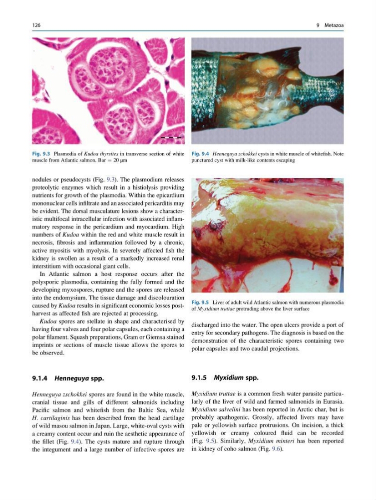
nodules or pseudocysts (Fig. 9.3). The plasmodium releases
proteolytic enzymes which result in a histiolysis providing
nutrients for growth of the plasmodia. Within the epicardium
mononuclear cells infiltrate and an associated pericarditis may
be evident. The dorsal musculature lesions show a characteristic multifocal intracellular infection with associated inflammatory response in the pericardium and myocardium. High
numbers of Kudoa within the red and white muscle result in
necrosis, fibrosis and inflammation followed by a chronic,
active myositis with myolysis. In severely affected fish the
kidney is swollen as a result of a markedly increased renal
interstitium with occasional giant cells.
In Atlantic salmon a host response occurs after the
polysporic plasmodia, containing the fully formed and the
developing myxospores, rupture and the spores are released
into the endomysium. The tissue damage and discolouration
caused by Kudoa results in significant economic losses postharvest as affected fish are rejected at processing.
Kudoa spores are stellate in shape and characterised by
having four valves and four polar capsules, each containing a
polar filament. Squash preparations, Gram or Giemsa stained
imprints or sections of muscle tissue allows the spores to
be observed.
9.1.4 Henneguya spp.
Henneguya zschokkei spores are found in the white muscle,
cranial tissue and gills of different salmonids including
Pacific salmon and whitefish from the Baltic Sea, while
H. cartilaginis has been described from the head cartilage
of wild masou salmon in Japan. Large, white-oval cysts with
a creamy content occur and ruin the aesthetic appearance of
the fillet (Fig. 9.4). The cysts mature and rupture through
the integument and a large number of infective spores are
discharged into the water. The open ulcers provide a port of
entry for secondary pathogens. The diagnosis is based on the
demonstration of the characteristic spores containing two
polar capsules and two caudal projections.
9.1.5 Myxidium spp.
Myxidium truttae is a common fresh water parasite particularly of the liver of wild and farmed salmonids in Eurasia.
Myxidium salvelini has been reported in Arctic char, but is
probably apathogenic. Grossly, affected livers may have
pale or yellowish surface protrusions. On incision, a thick
yellowish or creamy coloured fluid can be recorded
(Fig. 9.5). Similarly, Myxidium minteri has been reported
in kidney of coho salmon (Fig. 9.6).
Fig. 9.3 Plasmodia of Kudoa thyrsites in transverse section of white
muscle from Atlantic salmon. Bar ¼ 20 μm
Fig. 9.4 Henneguya zchokkei cysts in white muscle of whitefish. Note
punctured cyst with milk-like contents escaping
Fig. 9.5 Liver of adult wild Atlantic salmon with numerous plasmodia
of Myxidium truttae protruding above the liver surface
126
9 Metazoa
proteolytic enzymes which result in a histiolysis providing
nutrients for growth of the plasmodia. Within the epicardium
mononuclear cells infiltrate and an associated pericarditis may
be evident. The dorsal musculature lesions show a characteristic multifocal intracellular infection with associated inflammatory response in the pericardium and myocardium. High
numbers of Kudoa within the red and white muscle result in
necrosis, fibrosis and inflammation followed by a chronic,
active myositis with myolysis. In severely affected fish the
kidney is swollen as a result of a markedly increased renal
interstitium with occasional giant cells.
In Atlantic salmon a host response occurs after the
polysporic plasmodia, containing the fully formed and the
developing myxospores, rupture and the spores are released
into the endomysium. The tissue damage and discolouration
caused by Kudoa results in significant economic losses postharvest as affected fish are rejected at processing.
Kudoa spores are stellate in shape and characterised by
having four valves and four polar capsules, each containing a
polar filament. Squash preparations, Gram or Giemsa stained
imprints or sections of muscle tissue allows the spores to
be observed.
9.1.4 Henneguya spp.
Henneguya zschokkei spores are found in the white muscle,
cranial tissue and gills of different salmonids including
Pacific salmon and whitefish from the Baltic Sea, while
H. cartilaginis has been described from the head cartilage
of wild masou salmon in Japan. Large, white-oval cysts with
a creamy content occur and ruin the aesthetic appearance of
the fillet (Fig. 9.4). The cysts mature and rupture through
the integument and a large number of infective spores are
discharged into the water. The open ulcers provide a port of
entry for secondary pathogens. The diagnosis is based on the
demonstration of the characteristic spores containing two
polar capsules and two caudal projections.
9.1.5 Myxidium spp.
Myxidium truttae is a common fresh water parasite particularly of the liver of wild and farmed salmonids in Eurasia.
Myxidium salvelini has been reported in Arctic char, but is
probably apathogenic. Grossly, affected livers may have
pale or yellowish surface protrusions. On incision, a thick
yellowish or creamy coloured fluid can be recorded
(Fig. 9.5). Similarly, Myxidium minteri has been reported
in kidney of coho salmon (Fig. 9.6).
Fig. 9.3 Plasmodia of Kudoa thyrsites in transverse section of white
muscle from Atlantic salmon. Bar ¼ 20 μm
Fig. 9.4 Henneguya zchokkei cysts in white muscle of whitefish. Note
punctured cyst with milk-like contents escaping
Fig. 9.5 Liver of adult wild Atlantic salmon with numerous plasmodia
of Myxidium truttae protruding above the liver surface
126
9 Metazoa