

of the fungi from morphological characteristics and rDNA
ITS sequence data.
7.3
Phialophora sp.
Phialophora is both parasitic and saprophytic and described
as a systemic although infrequent infection of rainbow trout
and Atlantic salmon at low temperatures. In rainbow trout,
infection has been linked with cerebral mycetoma. Salmon
show fin haemorrhage and petechiae along the ventral surface. The internal organs are pale, the swim bladder filled
with a whitish, mucoid material and adherent to the body
wall and other internal organs, including the posterior
kidney and intestine. Apart from the haemorrhage in the
adipose tissue and mesentery, limited tissue response is
recorded to the hyphae. Microscopic examination reveals a
dense mycelium with septate and branching hyphae. The route
of infection is believed to be via the pneumatic duct when fry
fill the swim bladder with air from the surface. The source of
infection is unknown, but the genus Phialophora has a wide
distribution in the environment. In culture this fungus is slow
growing, with a thin-walled branching mycelium.
7.4
Phoma herbarum
Systemic infections of Phoma occur sporadically but
particularly in hatchery-reared Chinook salmon fingerlings
from the Pacific Northwest coast of America. Affected fish
show abnormal swimming behaviour, loss of equilibrium,
exophthalmia and sometimes haemorrhagic protrusion of the
vent. Internally, the most characteristic lesions comprise a
whitish creamy viscous mass in the swim bladder and
thickening of the wall, ascites and adhesions between visceral organs. Microscopically infection is associated with
necrosis and haemorrhage, with mild to moderate lymphocytic and histiocytic infiltration. Infection can spread to
other organs resulting in nephritis and a severe, systemic,
granulomatous reaction which includes the wall and lumen
of the dorsal aorta (Fig. 7.13).
Phoma spp., are saprophytic on a wide range of organic
material, including soil, plants and sewage. Infection is
believed to be via the pneumatic duct, however the absence
of pycnidia indicates that fish are not a natural site for
development. On Sabouraud medium with added dextrose
growth occurs as brownish colonies and microscopically
broad septate-branched hyphae that are hyaline, and
5–12 μm in diameter are reported. Diagnosis is based on
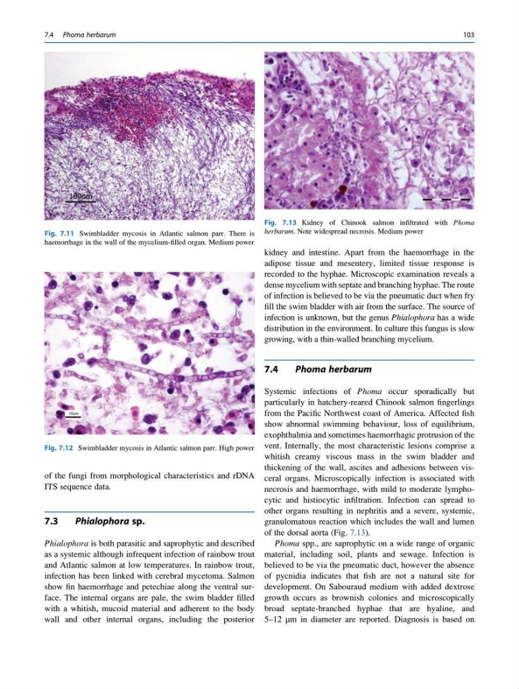
Fig. 7.11 Swimbladder mycosis in Atlantic salmon parr. There is
haemorrhage in the wall of the mycelium-filled organ. Medium power
Fig. 7.12 Swimbladder mycosis in Atlantic salmon parr. High power
Fig. 7.13 Kidney of Chinook salmon infiltrated with Phoma
herbarum. Note widespread necrosis. Medium power
7.4 Phoma herbarum
103
ITS sequence data.
7.3
Phialophora sp.
Phialophora is both parasitic and saprophytic and described
as a systemic although infrequent infection of rainbow trout
and Atlantic salmon at low temperatures. In rainbow trout,
infection has been linked with cerebral mycetoma. Salmon
show fin haemorrhage and petechiae along the ventral surface. The internal organs are pale, the swim bladder filled
with a whitish, mucoid material and adherent to the body
wall and other internal organs, including the posterior
kidney and intestine. Apart from the haemorrhage in the
adipose tissue and mesentery, limited tissue response is
recorded to the hyphae. Microscopic examination reveals a
dense mycelium with septate and branching hyphae. The route
of infection is believed to be via the pneumatic duct when fry
fill the swim bladder with air from the surface. The source of
infection is unknown, but the genus Phialophora has a wide
distribution in the environment. In culture this fungus is slow
growing, with a thin-walled branching mycelium.
7.4
Phoma herbarum
Systemic infections of Phoma occur sporadically but
particularly in hatchery-reared Chinook salmon fingerlings
from the Pacific Northwest coast of America. Affected fish
show abnormal swimming behaviour, loss of equilibrium,
exophthalmia and sometimes haemorrhagic protrusion of the
vent. Internally, the most characteristic lesions comprise a
whitish creamy viscous mass in the swim bladder and
thickening of the wall, ascites and adhesions between visceral organs. Microscopically infection is associated with
necrosis and haemorrhage, with mild to moderate lymphocytic and histiocytic infiltration. Infection can spread to
other organs resulting in nephritis and a severe, systemic,
granulomatous reaction which includes the wall and lumen
of the dorsal aorta (Fig. 7.13).
Phoma spp., are saprophytic on a wide range of organic
material, including soil, plants and sewage. Infection is
believed to be via the pneumatic duct, however the absence
of pycnidia indicates that fish are not a natural site for
development. On Sabouraud medium with added dextrose
growth occurs as brownish colonies and microscopically
broad septate-branched hyphae that are hyaline, and
5–12 μm in diameter are reported. Diagnosis is based on
Fig. 7.11 Swimbladder mycosis in Atlantic salmon parr. There is
haemorrhage in the wall of the mycelium-filled organ. Medium power
Fig. 7.12 Swimbladder mycosis in Atlantic salmon parr. High power
Fig. 7.13 Kidney of Chinook salmon infiltrated with Phoma
herbarum. Note widespread necrosis. Medium power
7.4 Phoma herbarum
103