

morphological characterization, the histological demonstration of PAS-positive hyphae in affected organs which can be
supported through molecular tools.
7.5
Exophiala spp.
Exophiala salmonis is an anamorphic black yeast, a fungus
from the family Herpotrichiellaceae. This species causes a low
prevalence internal systemic mycosis of marine-reared
salmonids e.g. Atlantic salmon. Infected fish may continue to
feed normally, but display erratic swimming movements which
may be followed by whirling behaviour. Exophthalmia and
cranial cutaneous ulcers are common, although these clinical
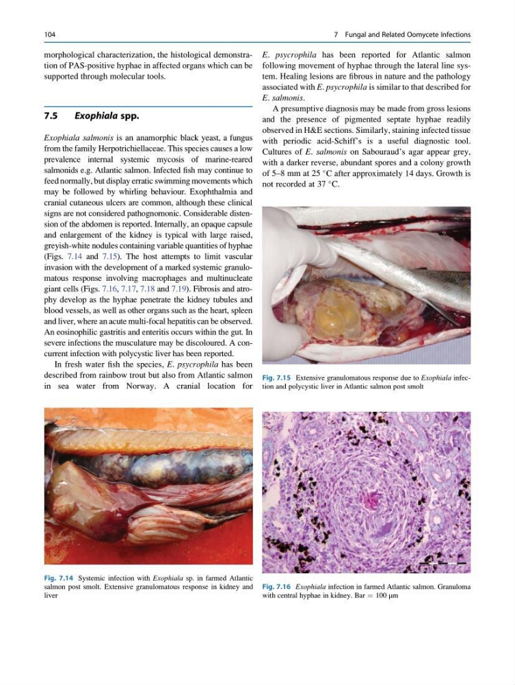
signs are not considered pathognomonic. Considerable distension of the abdomen is reported. Internally, an opaque capsule
and enlargement of the kidney is typical with large raised,
greyish-white nodules containing variable quantities of hyphae
(Figs. 7.14 and 7.15). The host attempts to limit vascular
invasion with the development of a marked systemic granulomatous response involving macrophages and multinucleate
giant cells (Figs. 7.16, 7.17, 7.18 and 7.19). Fibrosis and atrophy develop as the hyphae penetrate the kidney tubules and
blood vessels, as well as other organs such as the heart, spleen
and liver, where an acute multi-focal hepatitis can be observed.
An eosinophilic gastritis and enteritis occurs within the gut. In
severe infections the musculature may be discoloured. A concurrent infection with polycystic liver has been reported.
In fresh water fish the species, E. psycrophila has been
described from rainbow trout but also from Atlantic salmon
in sea water from Norway. A cranial location for
E. psycrophila has been reported for Atlantic salmon
following movement of hyphae through the lateral line system. Healing lesions are fibrous in nature and the pathology
associated with E. psycrophila is similar to that described for
E. salmonis.
A presumptive diagnosis may be made from gross lesions
and the presence of pigmented septate hyphae readily
observed in H&E sections. Similarly, staining infected tissue
with periodic acid-Schiff’s is a useful diagnostic tool.
Cultures of E. salmonis on Sabouraud’s agar appear grey,
with a darker reverse, abundant spores and a colony growth
of 5–8 mm at 25
C after approximately 14 days. Growth is
not recorded at 37
C.
Fig. 7.15 Extensive granulomatous response due to Exophiala infection and polycystic liver in Atlantic salmon post smolt
Fig. 7.14 Systemic infection with Exophiala sp. in farmed Atlantic
salmon post smolt. Extensive granulomatous response in kidney and
liver
Fig. 7.16 Exophiala infection in farmed Atlantic salmon. Granuloma
with central hyphae in kidney. Bar ¼ 100 μm
104
7 Fungal and Related Oomycete Infections
supported through molecular tools.
7.5
Exophiala spp.
Exophiala salmonis is an anamorphic black yeast, a fungus
from the family Herpotrichiellaceae. This species causes a low
prevalence internal systemic mycosis of marine-reared
salmonids e.g. Atlantic salmon. Infected fish may continue to
feed normally, but display erratic swimming movements which
may be followed by whirling behaviour. Exophthalmia and
cranial cutaneous ulcers are common, although these clinical
signs are not considered pathognomonic. Considerable distension of the abdomen is reported. Internally, an opaque capsule
and enlargement of the kidney is typical with large raised,
greyish-white nodules containing variable quantities of hyphae
(Figs. 7.14 and 7.15). The host attempts to limit vascular
invasion with the development of a marked systemic granulomatous response involving macrophages and multinucleate
giant cells (Figs. 7.16, 7.17, 7.18 and 7.19). Fibrosis and atrophy develop as the hyphae penetrate the kidney tubules and
blood vessels, as well as other organs such as the heart, spleen
and liver, where an acute multi-focal hepatitis can be observed.
An eosinophilic gastritis and enteritis occurs within the gut. In
severe infections the musculature may be discoloured. A concurrent infection with polycystic liver has been reported.
In fresh water fish the species, E. psycrophila has been
described from rainbow trout but also from Atlantic salmon
in sea water from Norway. A cranial location for
E. psycrophila has been reported for Atlantic salmon
following movement of hyphae through the lateral line system. Healing lesions are fibrous in nature and the pathology
associated with E. psycrophila is similar to that described for
E. salmonis.
A presumptive diagnosis may be made from gross lesions
and the presence of pigmented septate hyphae readily
observed in H&E sections. Similarly, staining infected tissue
with periodic acid-Schiff’s is a useful diagnostic tool.
Cultures of E. salmonis on Sabouraud’s agar appear grey,
with a darker reverse, abundant spores and a colony growth
of 5–8 mm at 25
C after approximately 14 days. Growth is
not recorded at 37
C.
Fig. 7.15 Extensive granulomatous response due to Exophiala infection and polycystic liver in Atlantic salmon post smolt
Fig. 7.14 Systemic infection with Exophiala sp. in farmed Atlantic
salmon post smolt. Extensive granulomatous response in kidney and
liver
Fig. 7.16 Exophiala infection in farmed Atlantic salmon. Granuloma
with central hyphae in kidney. Bar ¼ 100 μm
104
7 Fungal and Related Oomycete Infections