

clinical disease regressing at higher temperatures. Morbidity
can be high but RMS is usually a non-lethal condition where
appetite and growth are unaffected. Severe lesions may,
however, adversely affect the welfare of the fish.
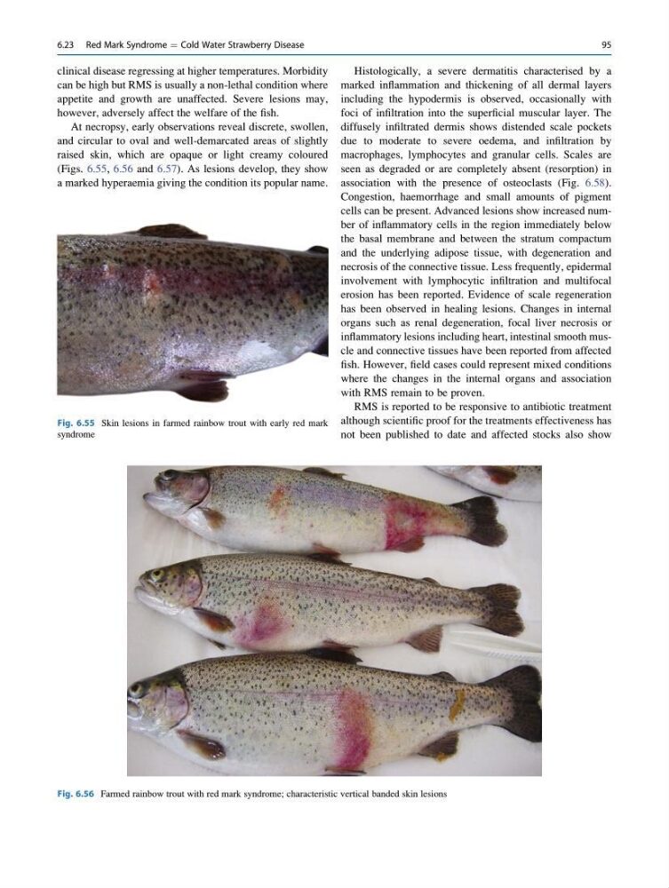
At necropsy, early observations reveal discrete, swollen,
and circular to oval and well-demarcated areas of slightly
raised skin, which are opaque or light creamy coloured
(Figs. 6.55, 6.56 and 6.57). As lesions develop, they show
a marked hyperaemia giving the condition its popular name.
Histologically, a severe dermatitis characterised by a
marked inflammation and thickening of all dermal layers
including the hypodermis is observed, occasionally with
foci of infiltration into the superficial muscular layer. The
diffusely infiltrated dermis shows distended scale pockets
due to moderate to severe oedema, and infiltration by
macrophages, lymphocytes and granular cells. Scales are
seen as degraded or are completely absent (resorption) in
association with the presence of osteoclasts (Fig. 6.58).
Congestion, haemorrhage and small amounts of pigment
cells can be present. Advanced lesions show increased number of inflammatory cells in the region immediately below
the basal membrane and between the stratum compactum
and the underlying adipose tissue, with degeneration and
necrosis of the connective tissue. Less frequently, epidermal
involvement with lymphocytic infiltration and multifocal
erosion has been reported. Evidence of scale regeneration
has been observed in healing lesions. Changes in internal
organs such as renal degeneration, focal liver necrosis or
inflammatory lesions including heart, intestinal smooth muscle and connective tissues have been reported from affected
fish. However, field cases could represent mixed conditions
where the changes in the internal organs and association
with RMS remain to be proven.
RMS is reported to be responsive to antibiotic treatment
although scientific proof for the treatments effectiveness has
not been published to date and affected stocks also show
Fig. 6.55 Skin lesions in farmed rainbow trout with early red mark
syndrome
Fig. 6.56 Farmed rainbow trout with red mark syndrome; characteristic vertical banded skin lesions
6.23 Red Mark Syndrome ¼ Cold Water Strawberry Disease
95
can be high but RMS is usually a non-lethal condition where
appetite and growth are unaffected. Severe lesions may,
however, adversely affect the welfare of the fish.
At necropsy, early observations reveal discrete, swollen,
and circular to oval and well-demarcated areas of slightly
raised skin, which are opaque or light creamy coloured
(Figs. 6.55, 6.56 and 6.57). As lesions develop, they show
a marked hyperaemia giving the condition its popular name.
Histologically, a severe dermatitis characterised by a
marked inflammation and thickening of all dermal layers
including the hypodermis is observed, occasionally with
foci of infiltration into the superficial muscular layer. The
diffusely infiltrated dermis shows distended scale pockets
due to moderate to severe oedema, and infiltration by
macrophages, lymphocytes and granular cells. Scales are
seen as degraded or are completely absent (resorption) in
association with the presence of osteoclasts (Fig. 6.58).
Congestion, haemorrhage and small amounts of pigment
cells can be present. Advanced lesions show increased number of inflammatory cells in the region immediately below
the basal membrane and between the stratum compactum
and the underlying adipose tissue, with degeneration and
necrosis of the connective tissue. Less frequently, epidermal
involvement with lymphocytic infiltration and multifocal
erosion has been reported. Evidence of scale regeneration
has been observed in healing lesions. Changes in internal
organs such as renal degeneration, focal liver necrosis or
inflammatory lesions including heart, intestinal smooth muscle and connective tissues have been reported from affected
fish. However, field cases could represent mixed conditions
where the changes in the internal organs and association
with RMS remain to be proven.
RMS is reported to be responsive to antibiotic treatment
although scientific proof for the treatments effectiveness has
not been published to date and affected stocks also show
Fig. 6.55 Skin lesions in farmed rainbow trout with early red mark
syndrome
Fig. 6.56 Farmed rainbow trout with red mark syndrome; characteristic vertical banded skin lesions
6.23 Red Mark Syndrome ¼ Cold Water Strawberry Disease
95