

Antarctic Fish Immunoglobulins: Structure and Antibody Specificity
181
A
B
PdSP
PdSP
COSP PdRP PdSP
; .
•
- 91
- 91
- 65
- 51
-I· - 9
- 65
- 47
- 51
- 40
54
. .
- .9
:
47
. , .t :...1 .
- 40
- 35
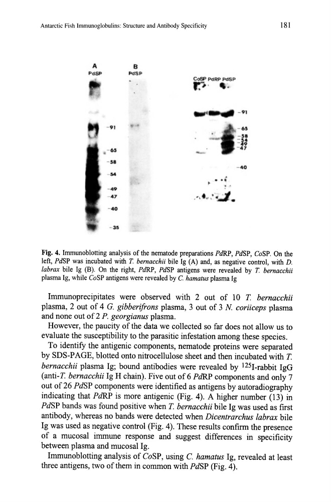
Fig. 4. Immunoblotting analysis of the nematode preparations PdRP, PdSP, CoSP. On the
left, PdSP was incubated with T. bernacchii bile Ig (A) and, as negative control, with D.
labrax bile Ig (B). On the right, PdRP, PdSP antigens were revealed by T. bernacchii
plasma Ig, while CoSP antigens were revealed by C. hamatus plasma Ig
Immunoprecipitates were observed with 2 out of lOT. bernacchii
plasma, 2 out of 4 G. gibberifrons plasma, 3 out of 3 N coriiceps plasma
and none out of 2 P. georgian us plasma.
However, the paucity of the data we collected so far does not allow us to
evaluate the susceptibility to the parasitic infestation among these species.
To identify the antigenic components, nematode proteins were separated
by SDS-PAGE, blotted onto nitrocellulose sheet and then incubated with T.
bernacchii plasma Ig; bound antibodies were revealed by l25I-rabbit IgG
(anti-T. bernacchii Ig H chain). Five out of 6 PdRP components and only 7
out of 26 PdSP components were identified as antigens by autoradiography
indicating that PdRP is more antigenic (Fig. 4). A higher number (13) in
PdSP bands was found positive when T. bernacchii bile Ig was used as first
antibody, whereas no bands were detected when Dicentrarchus labrax bile
Ig was used as negative control (Fig. 4). These results confirm the presence
of a mucosal immune response and suggest differences in specificity
between plasma and mucosal Ig.
Immunoblotting analysis of CoSP, using C. hamatus Ig, revealed at least
three antigens, two of them in common with PdSP (Fig. 4).
181
A
B
PdSP
PdSP
COSP PdRP PdSP
; .
•
- 91
- 91
- 65
- 51
-I· - 9
- 65
- 47
- 51
- 40
54
. .
- .9
:
47
. , .t :...1 .
- 40
- 35
Fig. 4. Immunoblotting analysis of the nematode preparations PdRP, PdSP, CoSP. On the
left, PdSP was incubated with T. bernacchii bile Ig (A) and, as negative control, with D.
labrax bile Ig (B). On the right, PdRP, PdSP antigens were revealed by T. bernacchii
plasma Ig, while CoSP antigens were revealed by C. hamatus plasma Ig
Immunoprecipitates were observed with 2 out of lOT. bernacchii
plasma, 2 out of 4 G. gibberifrons plasma, 3 out of 3 N coriiceps plasma
and none out of 2 P. georgian us plasma.
However, the paucity of the data we collected so far does not allow us to
evaluate the susceptibility to the parasitic infestation among these species.
To identify the antigenic components, nematode proteins were separated
by SDS-PAGE, blotted onto nitrocellulose sheet and then incubated with T.
bernacchii plasma Ig; bound antibodies were revealed by l25I-rabbit IgG
(anti-T. bernacchii Ig H chain). Five out of 6 PdRP components and only 7
out of 26 PdSP components were identified as antigens by autoradiography
indicating that PdRP is more antigenic (Fig. 4). A higher number (13) in
PdSP bands was found positive when T. bernacchii bile Ig was used as first
antibody, whereas no bands were detected when Dicentrarchus labrax bile
Ig was used as negative control (Fig. 4). These results confirm the presence
of a mucosal immune response and suggest differences in specificity
between plasma and mucosal Ig.
Immunoblotting analysis of CoSP, using C. hamatus Ig, revealed at least
three antigens, two of them in common with PdSP (Fig. 4).