

78
Fig.3.8. Haliclona oculata. Morphological skeleton constructed within the
contour shown in Fig.3.7f
Fig.3.9a,b. Haliclona oculata. Construction of the maximum discs (a) (solid
discs) and (b) (open discs), the branching rate rb, the branching angle b_angle,
and the geotropy angle g_angle within
the contour shown in Fig.3.7f
3. MEASURING GROWTH AND FORM
movement. For this species only a qualitative description of the amount of
exposure to water movement was available, where Class I indicates the lowest
and 11 the highest exposure to water movement.
For Haliclona oculata and Millepora alcicornis photographs of the samples were made using a high-contrast film in order to obtain sharp contours
(Kaandorp 1999). In the case of Pocillopora damicornis the original film
provided by Veron and Pichon was used, and sharp contour prints were
developed. The photographs were scanned using a 600 dpi scanner and converted into pbm-files: a lattice representation of the photograph where parts
of the object are in state "I " and the environment is in state "0". In Fig. 3.7
representative examples are shown of the resulting contour images of the
extreme growth forms of each of the three species.
3.3.2 Morphological Measurements in a Range of Growth Forms
The measurements are based primarily on the morphological skeleton of
the contour images. The morphological skeleton was obtained by applying
the thinning algorithm developed by Zhang and Suen (1984). The skeleton
is defined by connecting the center points of the maximum discs which fit
exactly within the contour (Rosenfeld and Kak 1982). In Fig. 3.8an example is
shown of the morphological skeleton constructed within the contour shown
in Fig.3.7f.ln the construction of the morphological skeleton several artefacts
may be generated. By occlusion effects some branches of the growth form
may overlap other branches and produce junctions that are not actually
present; furthermore, contamination and damage at the object (for example
at the holdfast of the organism) may produce "false" junctions. In many
marine sessile organisms (for example in many sponges) real junctions are
also present; these are formed by fusion of branches (anastomosis). In the
morphological measurements, both "false" junctions and junctions formed
by anastomosis were detected by comparing the images to the actual objects
and are not used in the measurements.
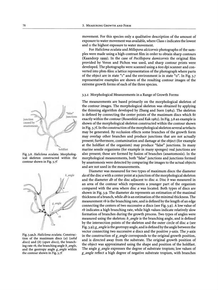
Diameter was measured for two types of maximum discs: the diameter
da of the disc a with a center point at a junction of the morphological skeleton
and the diameter db of the disc adjacent to disc a. Disc b was measured in
an area of the contour which represents a younger part of the organism
compared with the area where disc a was located. Both types of discs are
shown in Fig. 3.9. The diameter da represents an estimation of the maximal
thickness of a branch, while db is an estimation of the minimal thickness. The
measurement rb is the branching rate, and is defined by the length of an edge
connecting the centers of two successive a discs (see Fig.3.9). A low value of
rb indicates a high branching rate, while high values indicate relatively slow
formation of branches during the growth process . Two types of angles were
measured using the skeleton: b_angle is the branching angle, and is defined
by the intersection points of the skeleton and the outer circle of disc a (see
Fig. 3.9);g_angle is the geotropy angle, and is defined by the angle between the
vector connecting two successive a discs and the positive y-axis . The y-axis
in the construction of g_angle corresponds to the original growth position,
and is directed away from the substrate. The original growth position of
the object was approximated using the shape and position of the holdfast.
The angle g_angle expresses the degree of substrate tropism; low values of
g_angle reflect a high degree of negative substrate tropism, with branches
Fig.3.8. Haliclona oculata. Morphological skeleton constructed within the
contour shown in Fig.3.7f
Fig.3.9a,b. Haliclona oculata. Construction of the maximum discs (a) (solid
discs) and (b) (open discs), the branching rate rb, the branching angle b_angle,
and the geotropy angle g_angle within
the contour shown in Fig.3.7f
3. MEASURING GROWTH AND FORM
movement. For this species only a qualitative description of the amount of
exposure to water movement was available, where Class I indicates the lowest
and 11 the highest exposure to water movement.
For Haliclona oculata and Millepora alcicornis photographs of the samples were made using a high-contrast film in order to obtain sharp contours
(Kaandorp 1999). In the case of Pocillopora damicornis the original film
provided by Veron and Pichon was used, and sharp contour prints were
developed. The photographs were scanned using a 600 dpi scanner and converted into pbm-files: a lattice representation of the photograph where parts
of the object are in state "I " and the environment is in state "0". In Fig. 3.7
representative examples are shown of the resulting contour images of the
extreme growth forms of each of the three species.
3.3.2 Morphological Measurements in a Range of Growth Forms
The measurements are based primarily on the morphological skeleton of
the contour images. The morphological skeleton was obtained by applying
the thinning algorithm developed by Zhang and Suen (1984). The skeleton
is defined by connecting the center points of the maximum discs which fit
exactly within the contour (Rosenfeld and Kak 1982). In Fig. 3.8an example is
shown of the morphological skeleton constructed within the contour shown
in Fig.3.7f.ln the construction of the morphological skeleton several artefacts
may be generated. By occlusion effects some branches of the growth form
may overlap other branches and produce junctions that are not actually
present; furthermore, contamination and damage at the object (for example
at the holdfast of the organism) may produce "false" junctions. In many
marine sessile organisms (for example in many sponges) real junctions are
also present; these are formed by fusion of branches (anastomosis). In the
morphological measurements, both "false" junctions and junctions formed
by anastomosis were detected by comparing the images to the actual objects
and are not used in the measurements.
Diameter was measured for two types of maximum discs: the diameter
da of the disc a with a center point at a junction of the morphological skeleton
and the diameter db of the disc adjacent to disc a. Disc b was measured in
an area of the contour which represents a younger part of the organism
compared with the area where disc a was located. Both types of discs are
shown in Fig. 3.9. The diameter da represents an estimation of the maximal
thickness of a branch, while db is an estimation of the minimal thickness. The
measurement rb is the branching rate, and is defined by the length of an edge
connecting the centers of two successive a discs (see Fig.3.9). A low value of
rb indicates a high branching rate, while high values indicate relatively slow
formation of branches during the growth process . Two types of angles were
measured using the skeleton: b_angle is the branching angle, and is defined
by the intersection points of the skeleton and the outer circle of disc a (see
Fig. 3.9);g_angle is the geotropy angle, and is defined by the angle between the
vector connecting two successive a discs and the positive y-axis . The y-axis
in the construction of g_angle corresponds to the original growth position,
and is directed away from the substrate. The original growth position of
the object was approximated using the shape and position of the holdfast.
The angle g_angle expresses the degree of substrate tropism; low values of
g_angle reflect a high degree of negative substrate tropism, with branches