

44
(a)
(b)
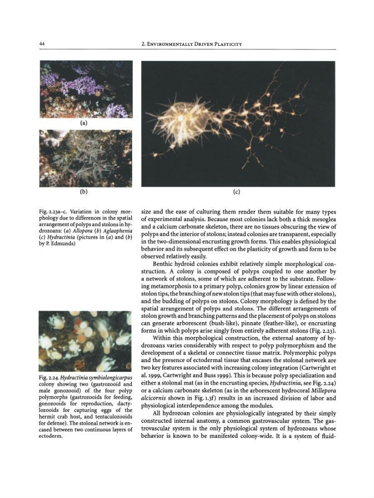
Fig.z.zja-c. Variation in colony morphology due to differences in the spatial
arrangement ofpolypsand stolons in hydrozoans: (a) Allopora (b) Aglaophenia
(c) Hydraetinia (pictures in (a) and (b)
by P. Edmunds)
Fig. 2.24. Hydractiniasymbiolongicarpus
colony showing two (gastrozooid and
male gonozooid) of the four polyp
polymorphs (gastrozooids for feeding,
gonozooids for reproduction, dactylozooids for capturing eggs of the
hermit crab host, and tentaculozooids
for defense). The stolonal network is encased between two continuous layers of
ectoderm.
2. ENVIRONMENTALLY DRIVEN PLASTICITY
(c)
size and the ease of culturing them render them suitable for many types
of experimental analysis. Because most colonies lack both a thick mesoglea
and a calcium carbonate skeleton, there are no tissues obscuring the view of
polyps and the interior of stolons; instead colonies are transparent, especially
in the two-dimensional encrusting growth forms. This enables physiological
behavior and its subsequent effect on the plasticity of growth and form to be
observed relatively easily.
Benthic hydroid colonies exhibit relatively simple morphological construction. A colony is composed of polyps coupled to one another by
a network of stolons, some of which are adherent to the substrate. Following metamorphosis to a primary polyp, colonies grow by linear extension of
stolon tips, the branching of new stolon tips (that may fuse with other stolons),
and the budding of polyps on stolons. Colony morphology is defined by the
spatial arrangement of polyps and stolons. The different arrangements of
stolon growth and branching patterns and the placement of polyps on stolons
can generate arborescent (bush-like), pinnate (feather-like), or encrusting
forms in which polyps arise singly from entirely adherent stolons (Fig. 2.23).
Within this morphological construction, the external anatomy of hydrozoans varies considerably with respect to polyp polymorphism and the
development of a skeletal or connective tissue matrix. Polymorphic polyps
and the presence of ectodermal tissue that encases the stolonal network are
two key features associated with increasing colony integration (Cartwright et
al. 1999, Cartwright and Buss 1999). This is because polyp specialization and
either a stolonal mat (as in the encrusting species, Hydractinia, see Fig. 2.24)
or a calcium carbonate skeleton (as in the arborescent hydrocoral Millepora
alcicornis shown in Pig.r.jf) results in an increased division of labor and
physiological interdependence among the modules.
All hydrozoan colonies are physiologically integrated by their simply
constructed internal anatomy, a common gastrovascular system. The gastrovascular system is the only physiological system of hydrozoans whose
behavior is known to be manifested colony-wide. It is a system of fluid-
(a)
(b)
Fig.z.zja-c. Variation in colony morphology due to differences in the spatial
arrangement ofpolypsand stolons in hydrozoans: (a) Allopora (b) Aglaophenia
(c) Hydraetinia (pictures in (a) and (b)
by P. Edmunds)
Fig. 2.24. Hydractiniasymbiolongicarpus
colony showing two (gastrozooid and
male gonozooid) of the four polyp
polymorphs (gastrozooids for feeding,
gonozooids for reproduction, dactylozooids for capturing eggs of the
hermit crab host, and tentaculozooids
for defense). The stolonal network is encased between two continuous layers of
ectoderm.
2. ENVIRONMENTALLY DRIVEN PLASTICITY
(c)
size and the ease of culturing them render them suitable for many types
of experimental analysis. Because most colonies lack both a thick mesoglea
and a calcium carbonate skeleton, there are no tissues obscuring the view of
polyps and the interior of stolons; instead colonies are transparent, especially
in the two-dimensional encrusting growth forms. This enables physiological
behavior and its subsequent effect on the plasticity of growth and form to be
observed relatively easily.
Benthic hydroid colonies exhibit relatively simple morphological construction. A colony is composed of polyps coupled to one another by
a network of stolons, some of which are adherent to the substrate. Following metamorphosis to a primary polyp, colonies grow by linear extension of
stolon tips, the branching of new stolon tips (that may fuse with other stolons),
and the budding of polyps on stolons. Colony morphology is defined by the
spatial arrangement of polyps and stolons. The different arrangements of
stolon growth and branching patterns and the placement of polyps on stolons
can generate arborescent (bush-like), pinnate (feather-like), or encrusting
forms in which polyps arise singly from entirely adherent stolons (Fig. 2.23).
Within this morphological construction, the external anatomy of hydrozoans varies considerably with respect to polyp polymorphism and the
development of a skeletal or connective tissue matrix. Polymorphic polyps
and the presence of ectodermal tissue that encases the stolonal network are
two key features associated with increasing colony integration (Cartwright et
al. 1999, Cartwright and Buss 1999). This is because polyp specialization and
either a stolonal mat (as in the encrusting species, Hydractinia, see Fig. 2.24)
or a calcium carbonate skeleton (as in the arborescent hydrocoral Millepora
alcicornis shown in Pig.r.jf) results in an increased division of labor and
physiological interdependence among the modules.
All hydrozoan colonies are physiologically integrated by their simply
constructed internal anatomy, a common gastrovascular system. The gastrovascular system is the only physiological system of hydrozoans whose
behavior is known to be manifested colony-wide. It is a system of fluid-