

150
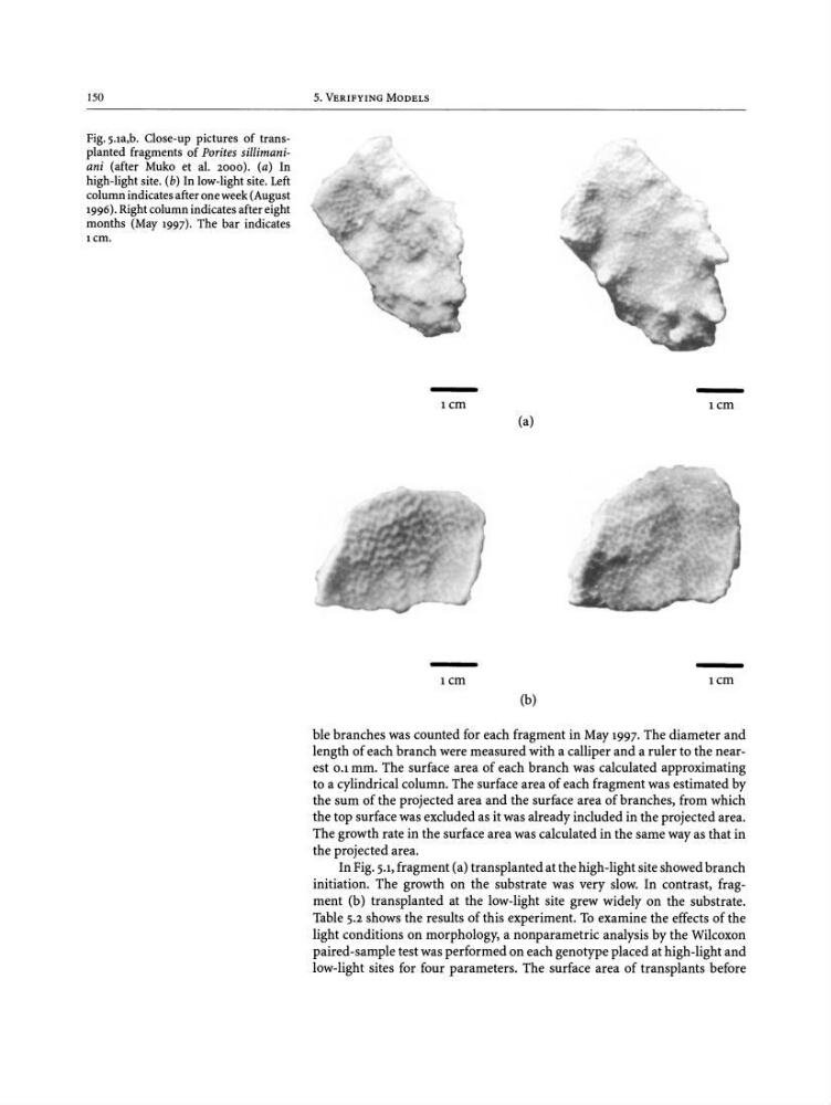
Pig.y .ra.b . Close-up pictures of transplanted fragments of Porites sillimaniani (after Muko et al. 2000) . (a) In
high-light site . (b) In low-light site. Left
column indicates after one week (August
1996) . Right column indicates after eight
months (May 1997). The bar indicates
i cm ,
5. VERIFYING MODELS
i cm
i cm
(a)
(b)
i cm
i cm
ble branches was counted for each fragment in May 1997. The diameter and
length of each branch were measured with a calliper and a ruler to the nearest 0 .1 rnm . The surface area of each branch was calculated approximating
to a cylindrical column. The surface area of each fragment was estimated by
the sum of the projected area and the surface area of branches, from which
the top surface was excluded as it was already included in the projected area .
The growth rate in the surface area was calculated in the same way as that in
the projected area .
In Fig. 5.1,fragment (a) transplanted at the high-light site showed branch
initiation. The growth on the substrate was very slow. In contrast, frag -
ment (b) transplanted at the low-light site grew widely on the substrate.
Table 5.2 shows the results of this experiment. To examine the effects of the
light conditions on morphology, a nonparametric analysis by the Wilcoxon
paired-sample test was performed on each genotype placed at high-light and
low-light sites for four parameters. The surface area of transplants before
Pig.y .ra.b . Close-up pictures of transplanted fragments of Porites sillimaniani (after Muko et al. 2000) . (a) In
high-light site . (b) In low-light site. Left
column indicates after one week (August
1996) . Right column indicates after eight
months (May 1997). The bar indicates
i cm ,
5. VERIFYING MODELS
i cm
i cm
(a)
(b)
i cm
i cm
ble branches was counted for each fragment in May 1997. The diameter and
length of each branch were measured with a calliper and a ruler to the nearest 0 .1 rnm . The surface area of each branch was calculated approximating
to a cylindrical column. The surface area of each fragment was estimated by
the sum of the projected area and the surface area of branches, from which
the top surface was excluded as it was already included in the projected area .
The growth rate in the surface area was calculated in the same way as that in
the projected area .
In Fig. 5.1,fragment (a) transplanted at the high-light site showed branch
initiation. The growth on the substrate was very slow. In contrast, frag -
ment (b) transplanted at the low-light site grew widely on the substrate.
Table 5.2 shows the results of this experiment. To examine the effects of the
light conditions on morphology, a nonparametric analysis by the Wilcoxon
paired-sample test was performed on each genotype placed at high-light and
low-light sites for four parameters. The surface area of transplants before