

5.1. TRANSPLANTATION EXPER IMENTS WITH STONY CORALS
this experiment. The details of translocation of photosynthetic products be -
tween the polyps are described in Rinkevich and Loya (1983b), Oren et al.
(1997) and in Sect. 2.2.4.
EXAMPLE OF PORITES SILLIMANIANI. In this section an example is given of
transplantation experiments with the stony coral Porites sillimaniani which
examine the plastic change of growth form in response to light conditions.
Porites sillimaniani is a common species in a variety of reef environments of
the tropical Pacific, and it displays a striking variation of the whole colony
morphology with respect to light availability (see Fig. 2.32). The transplantation experiment, in which clonal replicates are exposed to different light
conditions, can solve the problem of whether the morphological variation is
based on genetic differences or due to phenotypic plasticity. Details of this
transplantation experiment can be found in Muko et al. (2000).
Two transplant sites were established side by side within 3 m on the sea
bottom 6 m in depth in Okinawa, Japan. Each site had two iron frames in order
to manipulate the light intensity. The light intensity was controlled using
plexiglass covers. The other environmental factors, such as water velocity,
salinity, nutrient level, and so on, were kept unchanged. The irradiance on
the horizontal plane at the high-light site was significantly higher than that
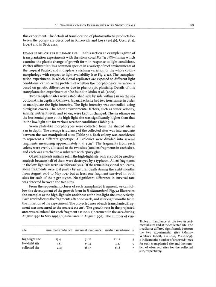
in the low-light site for various weather conditions (Table 5.1).
Seven plate-like morphotypes were collected from the shaded site at
4 m in depth. The average irradiance of the collected sites was intermediate
between the two manipulated sites (Table 5.1). Each colony was considered
to represent a different genotype. All colonies were divided into several
fragments measuring approximately 3 x 3 ern", The fragments from each
colony were evenly allocated to the two sites (total 26 fragments in each site),
and each was attached to a substrate with epoxy glue.
Of 26 fragments initially set in the high-light site, only 13 could be used for
analysis because half of them were destroyed by a typhoon. All 26 fragments
in the low-light site were used for analysis. Of the remaining clonal replicates,
some fragments were lost partly by natural death during the eight months
from August 1996 to May 1997 but at least one fragment survived in both
sites for each of the 7 genotypes. No significant difference in survival rate
was detected between the two sites .
From the sequential pictures of each transplanted fragment, we can follow the development of the growth form in P. sillimaniani. Fig. 5.1illustrates
the examples at the high-light site and those at the low-light site, respectively.
Each row indicates the fragments after one week, and after eight months from
the initiation of the experiment. The projected area of each transplanted fragment was measured to the nearest 0.1crrr'. The growth rate in the projected
area was calculated for each fragment as: 100 x (increment in the area during
August 1996 to May 1997) / (initial area in August 1996). The number of visi149
site
high-light site
low-light site
collectedsite
minimal irradiance
15·4
1.51
2·47
maximal irradiance
31.28
14·35
18.17
median irradiance n
22.12
5
3·22
5
6.18
7
Table 5.1. Irradiance at the two experimental sitesand at the collectedsite. The
irradiance differedsignificantly between
the two experimental sites (MannWhitney U-test, z = -2.6, P = 0.009).
n indicatesthe number ofobservedtimes
for each transplanted site and the number of observed sites for the collected
site, respectively.
this experiment. The details of translocation of photosynthetic products be -
tween the polyps are described in Rinkevich and Loya (1983b), Oren et al.
(1997) and in Sect. 2.2.4.
EXAMPLE OF PORITES SILLIMANIANI. In this section an example is given of
transplantation experiments with the stony coral Porites sillimaniani which
examine the plastic change of growth form in response to light conditions.
Porites sillimaniani is a common species in a variety of reef environments of
the tropical Pacific, and it displays a striking variation of the whole colony
morphology with respect to light availability (see Fig. 2.32). The transplantation experiment, in which clonal replicates are exposed to different light
conditions, can solve the problem of whether the morphological variation is
based on genetic differences or due to phenotypic plasticity. Details of this
transplantation experiment can be found in Muko et al. (2000).
Two transplant sites were established side by side within 3 m on the sea
bottom 6 m in depth in Okinawa, Japan. Each site had two iron frames in order
to manipulate the light intensity. The light intensity was controlled using
plexiglass covers. The other environmental factors, such as water velocity,
salinity, nutrient level, and so on, were kept unchanged. The irradiance on
the horizontal plane at the high-light site was significantly higher than that
in the low-light site for various weather conditions (Table 5.1).
Seven plate-like morphotypes were collected from the shaded site at
4 m in depth. The average irradiance of the collected sites was intermediate
between the two manipulated sites (Table 5.1). Each colony was considered
to represent a different genotype. All colonies were divided into several
fragments measuring approximately 3 x 3 ern", The fragments from each
colony were evenly allocated to the two sites (total 26 fragments in each site),
and each was attached to a substrate with epoxy glue.
Of 26 fragments initially set in the high-light site, only 13 could be used for
analysis because half of them were destroyed by a typhoon. All 26 fragments
in the low-light site were used for analysis. Of the remaining clonal replicates,
some fragments were lost partly by natural death during the eight months
from August 1996 to May 1997 but at least one fragment survived in both
sites for each of the 7 genotypes. No significant difference in survival rate
was detected between the two sites .
From the sequential pictures of each transplanted fragment, we can follow the development of the growth form in P. sillimaniani. Fig. 5.1illustrates
the examples at the high-light site and those at the low-light site, respectively.
Each row indicates the fragments after one week, and after eight months from
the initiation of the experiment. The projected area of each transplanted fragment was measured to the nearest 0.1crrr'. The growth rate in the projected
area was calculated for each fragment as: 100 x (increment in the area during
August 1996 to May 1997) / (initial area in August 1996). The number of visi149
site
high-light site
low-light site
collectedsite
minimal irradiance
15·4
1.51
2·47
maximal irradiance
31.28
14·35
18.17
median irradiance n
22.12
5
3·22
5
6.18
7
Table 5.1. Irradiance at the two experimental sitesand at the collectedsite. The
irradiance differedsignificantly between
the two experimental sites (MannWhitney U-test, z = -2.6, P = 0.009).
n indicatesthe number ofobservedtimes
for each transplanted site and the number of observed sites for the collected
site, respectively.