

17 Nematoda
205
solitary. The latter cell type exhibited cell division. Uniform spherical
granular cells, which developed fine cytoplasmic projections appeared
three months after initiation of the culture. These spherical granular cells
showed unequal division and formed a confluent monolayer, and could
be subcultured continuously (Fig. 2).
Subprotocol 2
Ovaries of Rat Parasitic Nematodes (Rhabditida)
The following is Ogura's method (1989) for culturing ovary cells of the
rat parasitic nematode Angiostrongylus cantonensis .
• • Materials
- CO 2 incubator
- Petri dish
- Maximov slide
- Surgical knife
- Forceps
- Needles
- Pasteur pipettes
- T -15 culture flasks
- 0.9% NaCI solution
- Ogura's medium (see Appendix 1)
- Silk thread
- Adult female Angiostrangylus cantonensis
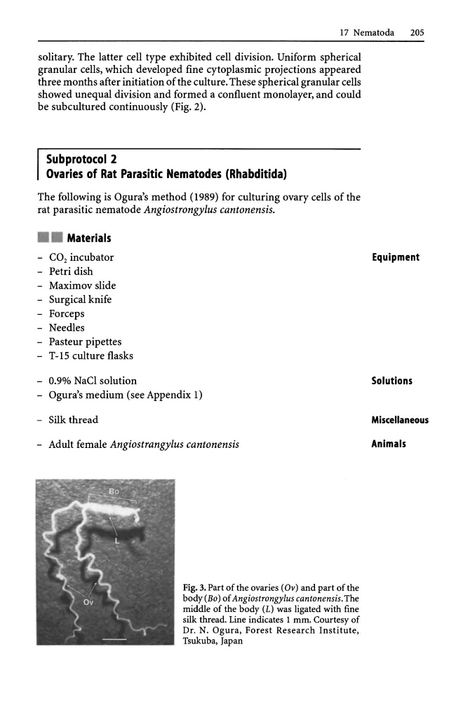
Fig. 3. Part of the ovaries (Ov) and part of the
body (Bo) of Angiostrongylus cantonensis. The
middle of the body (L) was ligated with fine
silk thread. Line indicates 1 mm. Courtesy of
Dr. N. Ogura, Forest Research Institute,
Tsukuba, Japan
Equipment
Solutions
Miscellaneous
Animals
205
solitary. The latter cell type exhibited cell division. Uniform spherical
granular cells, which developed fine cytoplasmic projections appeared
three months after initiation of the culture. These spherical granular cells
showed unequal division and formed a confluent monolayer, and could
be subcultured continuously (Fig. 2).
Subprotocol 2
Ovaries of Rat Parasitic Nematodes (Rhabditida)
The following is Ogura's method (1989) for culturing ovary cells of the
rat parasitic nematode Angiostrongylus cantonensis .
• • Materials
- CO 2 incubator
- Petri dish
- Maximov slide
- Surgical knife
- Forceps
- Needles
- Pasteur pipettes
- T -15 culture flasks
- 0.9% NaCI solution
- Ogura's medium (see Appendix 1)
- Silk thread
- Adult female Angiostrangylus cantonensis
Fig. 3. Part of the ovaries (Ov) and part of the
body (Bo) of Angiostrongylus cantonensis. The
middle of the body (L) was ligated with fine
silk thread. Line indicates 1 mm. Courtesy of
Dr. N. Ogura, Forest Research Institute,
Tsukuba, Japan
Equipment
Solutions
Miscellaneous
Animals