

206
Methods for Setting Up Primary Cultures Specific to Animal Groups
• • Procedure
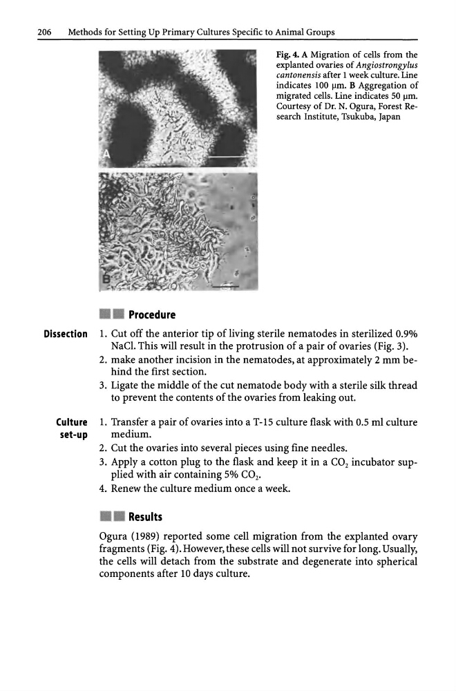
Fig. 4. A Migration of cells from the
explanted ovaries of Angiostrongylus
cantonensis after 1 week culture. Line
indicates 100 J..Im. B Aggregation of
migrated cells. Line indicates 50 J..Im.
Courtesy of Dr. N. Ogura, Forest Research Institute, Tsukuba, Japan
Dissection l. Cut off the anterior tip of living sterile nematodes in sterilized 0.9%
NaCl. This will result in the protrusion of a pair of ovaries (Fig. 3).
2. make another incision in the nematodes, at approximately 2 mm behind the first section.
3. Ligate the middle of the cut nematode body with a sterile silk thread
to prevent the contents of the ovaries from leaking out.
Culture l. Transfer a pair of ovaries into a T-15 culture flask with 0.5 ml culture
set-up
medium.
2. Cut the ovaries into several pieces using fine needles.
3. Apply a cotton plug to the flask and keep it in a CO 2 incubator supplied with air containing 5% CO 2 ,
4. Renew the culture medium once a week.
• • Results
Ogura (1989) reported some cell migration from the explanted ovary
fragments (Fig. 4). However, these cells will not survive for long. Usually,
the cells will detach from the substrate and degenerate into spherical
components after 10 days culture.
Methods for Setting Up Primary Cultures Specific to Animal Groups
• • Procedure
Fig. 4. A Migration of cells from the
explanted ovaries of Angiostrongylus
cantonensis after 1 week culture. Line
indicates 100 J..Im. B Aggregation of
migrated cells. Line indicates 50 J..Im.
Courtesy of Dr. N. Ogura, Forest Research Institute, Tsukuba, Japan
Dissection l. Cut off the anterior tip of living sterile nematodes in sterilized 0.9%
NaCl. This will result in the protrusion of a pair of ovaries (Fig. 3).
2. make another incision in the nematodes, at approximately 2 mm behind the first section.
3. Ligate the middle of the cut nematode body with a sterile silk thread
to prevent the contents of the ovaries from leaking out.
Culture l. Transfer a pair of ovaries into a T-15 culture flask with 0.5 ml culture
set-up
medium.
2. Cut the ovaries into several pieces using fine needles.
3. Apply a cotton plug to the flask and keep it in a CO 2 incubator supplied with air containing 5% CO 2 ,
4. Renew the culture medium once a week.
• • Results
Ogura (1989) reported some cell migration from the explanted ovary
fragments (Fig. 4). However, these cells will not survive for long. Usually,
the cells will detach from the substrate and degenerate into spherical
components after 10 days culture.