

192
Methods for Setting Up Primary Cultures Specific to Animal Groups
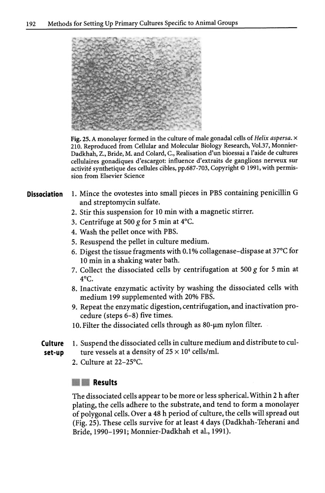
Fig. 25. A monolayer formed in the culture of male gonadal cells of Helix aspersa. x
210. Reproduced from Cellular and Molecular Biology Research, Vol.37, MonnierDadkhah, Z., Bride, M. and Colard, c., Realisation d'un bioessai a l'aide de cultures
cellulaires gonadiques d'escargot: influence d'extraits de ganglions nerveux sur
activite synthetique des cellules cibles, pp.687-703, Copyright © 1991, with permission from Elsevier Science
Dissociation 1. Mince the ovotestes into small pieces in PBS containing penicillin G
and streptomycin sulfate.
2. Stir this suspension for 10 min with a magnetic stirrer.
3. Centrifuge at 500 g for 5 min at 4°C.
4. Wash the pellet once with PBS.
5. Resuspend the pellet in culture medium.
6. Digest the tissue fragments with 0.1 % collagenase-dispase at 37°C for
10 min in a shaking water bath.
7. Collect the dissociated cells by centrifugation at 500 g for 5 min at
4°C.
8. Inactivate enzymatic activity by washing the dissociated cells with
medium 199 supplemented with 20% FBS.
9. Repeat the enzymatic digestion, centrifugation, and inactivation procedure (steps 6-8) five times.
10. Filter the dissociated cells through as 80-flm nylon fIlter.
Culture 1. Suspend the dissociated cells in culture medium and distribute to culset-up
ture vessels at a density of 25 x 10 4 cells/ml.
2. Culture at 22-25°C .
• • Results
The dissociated cells appear to be more or less spherical. Within 2 h after
plating, the cells adhere to the substrate, and tend to form a monolayer
of polygonal cells. Over a 48 h period of culture, the cells will spread out
(Fig. 25). These cells survive for at least 4 days (Dadkhah-Teherani and
Bride, 1990-1991; Monnier-Dadkhah et aI., 1991).
Methods for Setting Up Primary Cultures Specific to Animal Groups
Fig. 25. A monolayer formed in the culture of male gonadal cells of Helix aspersa. x
210. Reproduced from Cellular and Molecular Biology Research, Vol.37, MonnierDadkhah, Z., Bride, M. and Colard, c., Realisation d'un bioessai a l'aide de cultures
cellulaires gonadiques d'escargot: influence d'extraits de ganglions nerveux sur
activite synthetique des cellules cibles, pp.687-703, Copyright © 1991, with permission from Elsevier Science
Dissociation 1. Mince the ovotestes into small pieces in PBS containing penicillin G
and streptomycin sulfate.
2. Stir this suspension for 10 min with a magnetic stirrer.
3. Centrifuge at 500 g for 5 min at 4°C.
4. Wash the pellet once with PBS.
5. Resuspend the pellet in culture medium.
6. Digest the tissue fragments with 0.1 % collagenase-dispase at 37°C for
10 min in a shaking water bath.
7. Collect the dissociated cells by centrifugation at 500 g for 5 min at
4°C.
8. Inactivate enzymatic activity by washing the dissociated cells with
medium 199 supplemented with 20% FBS.
9. Repeat the enzymatic digestion, centrifugation, and inactivation procedure (steps 6-8) five times.
10. Filter the dissociated cells through as 80-flm nylon fIlter.
Culture 1. Suspend the dissociated cells in culture medium and distribute to culset-up
ture vessels at a density of 25 x 10 4 cells/ml.
2. Culture at 22-25°C .
• • Results
The dissociated cells appear to be more or less spherical. Within 2 h after
plating, the cells adhere to the substrate, and tend to form a monolayer
of polygonal cells. Over a 48 h period of culture, the cells will spread out
(Fig. 25). These cells survive for at least 4 days (Dadkhah-Teherani and
Bride, 1990-1991; Monnier-Dadkhah et aI., 1991).