

the y make a 'hairpin' bend and follow a parallel course back to the cortex, merging
into the distal tubule and the collecting ducts. Certain of the renal capillaries (vasa
recta) follow a similar pattern, and the two types of vessels together function as
a 'countercurrent multiplier' system, to create a hyperosmotic concentration
gradient which reaches a maximum at the 'hairpin bend'. Such a system allows birds
and mammals to form urine that is hyperosmotic to the plasma, and was first described by HARGITAY and KUHN (1951). An excellent description is given by
GOTTSCHALK and MYLLE (1959) (see Fig. 1.3). In 1944 SPERBER noted that mammals with long loops of HENLE and the elongated renal papillae associated with
this , concentrated their urine more than those with shorter loops. This is due to
the greater osmotic gradients which more elongated 'countercurrent' systems can
form .
The blood supply to the glomerulus is arterial, entering and leaving the capillary
tuft as afferent and efferent glomerular arterioles. It then breaks down into a second
network of peritubular capillaries and vasa recta, which supply the tubular tissue.
A renal portal system is present in birds, reptiles, Amphibia and most fishes (it
is not for instance present in lampreys). Venous blood from the posterior regions
of the body, passes into the renal portal vein from whence it may either supply
the tubules (but not the glomeruli), or be shunted by a more direct route into the
efferent renal vein (H. SMITH, 1951).
y) Physiology. The formation of urine involves the ultrafiltration of the plasma
through the glomerulus, reabsorption of solutes and water from the lumen of the
tubule, as well as secretion of materials by the tubular cells into the tubular fluid.
These processes have been studied by a variety of techniques, the most elegant involving collection and analysis of fluid from different regions of the nephron by
means of rnicropipettes.
a) Glomerular f iltration. Plasma is filtered in the glomerulus in the same manner
as in other capillaries. The filtrate is relatively free of proteins, but otherwise has
a similar composition to the plasma. Such ultrafiltration is caused by hydrostatic
forces in the arterial system, which are opposed in the nephron by the osmotic
pressure of the proteins (equivalent to 25 mm Hg in mammals but usually about
half this in other groups), the external tissue pressure on the tubule (10 mm Hg),
along with the back pressure of the system.
The glomerular filtration rate (GFR) is thought to be best reflected by the renal
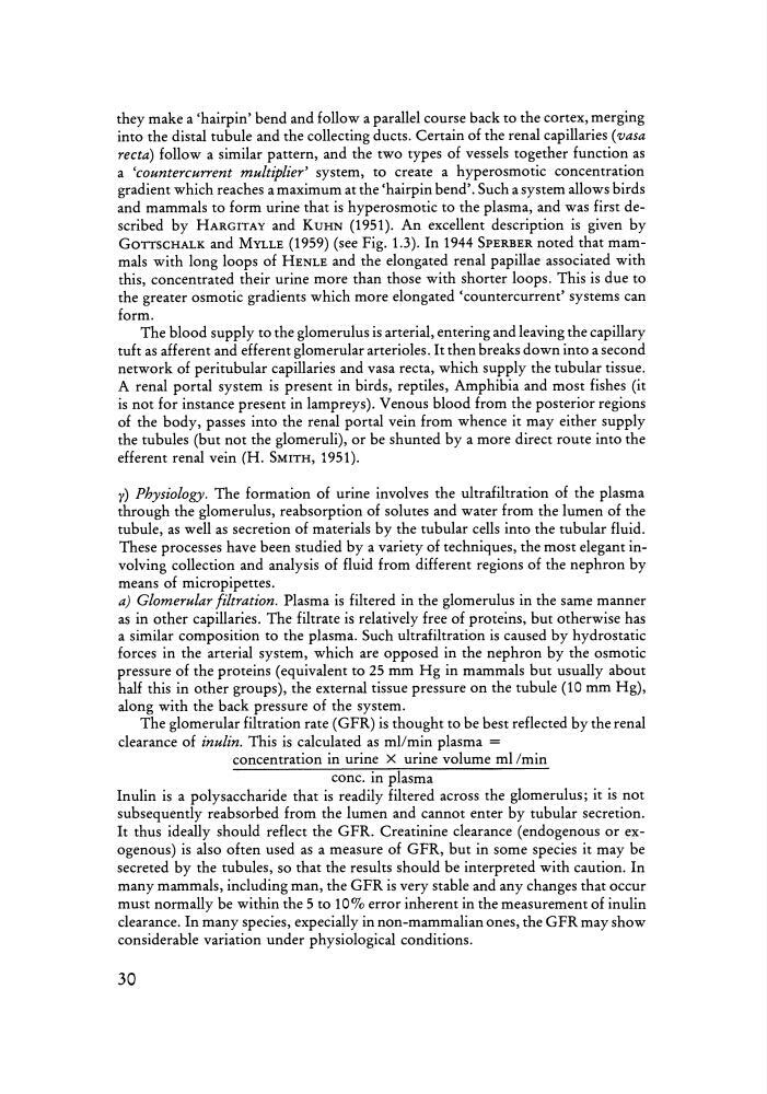
clearance of inulin. This is calculated as mllmin plasma =
concentration in urine X urine volume ml/min
cone. in plasma
Inulin is a polysaccharide that is readily filtered across the glomerulus; it is not
subsequently reabsorbed from the lumen and cannot enter by tubular secretion.
It thus ideall y should reflect the GFR. Creatinine clearance (endogenous or exogenous) is also often used as a measure of GFR, but in som e species it may be
secreted by the tubules, so that the results should be interpreted with caution. In
many mammals, including man, the GFR is very stable and any changes that occur
must normally be within the 5 to 10 % error inherent in the mea surement of inulin
clearance. In many species, expecially in non-mammalian ones, the GFR ma y show
considerable variatio n under ph ysiological conditions.
30
into the distal tubule and the collecting ducts. Certain of the renal capillaries (vasa
recta) follow a similar pattern, and the two types of vessels together function as
a 'countercurrent multiplier' system, to create a hyperosmotic concentration
gradient which reaches a maximum at the 'hairpin bend'. Such a system allows birds
and mammals to form urine that is hyperosmotic to the plasma, and was first described by HARGITAY and KUHN (1951). An excellent description is given by
GOTTSCHALK and MYLLE (1959) (see Fig. 1.3). In 1944 SPERBER noted that mammals with long loops of HENLE and the elongated renal papillae associated with
this , concentrated their urine more than those with shorter loops. This is due to
the greater osmotic gradients which more elongated 'countercurrent' systems can
form .
The blood supply to the glomerulus is arterial, entering and leaving the capillary
tuft as afferent and efferent glomerular arterioles. It then breaks down into a second
network of peritubular capillaries and vasa recta, which supply the tubular tissue.
A renal portal system is present in birds, reptiles, Amphibia and most fishes (it
is not for instance present in lampreys). Venous blood from the posterior regions
of the body, passes into the renal portal vein from whence it may either supply
the tubules (but not the glomeruli), or be shunted by a more direct route into the
efferent renal vein (H. SMITH, 1951).
y) Physiology. The formation of urine involves the ultrafiltration of the plasma
through the glomerulus, reabsorption of solutes and water from the lumen of the
tubule, as well as secretion of materials by the tubular cells into the tubular fluid.
These processes have been studied by a variety of techniques, the most elegant involving collection and analysis of fluid from different regions of the nephron by
means of rnicropipettes.
a) Glomerular f iltration. Plasma is filtered in the glomerulus in the same manner
as in other capillaries. The filtrate is relatively free of proteins, but otherwise has
a similar composition to the plasma. Such ultrafiltration is caused by hydrostatic
forces in the arterial system, which are opposed in the nephron by the osmotic
pressure of the proteins (equivalent to 25 mm Hg in mammals but usually about
half this in other groups), the external tissue pressure on the tubule (10 mm Hg),
along with the back pressure of the system.
The glomerular filtration rate (GFR) is thought to be best reflected by the renal
clearance of inulin. This is calculated as mllmin plasma =
concentration in urine X urine volume ml/min
cone. in plasma
Inulin is a polysaccharide that is readily filtered across the glomerulus; it is not
subsequently reabsorbed from the lumen and cannot enter by tubular secretion.
It thus ideall y should reflect the GFR. Creatinine clearance (endogenous or exogenous) is also often used as a measure of GFR, but in som e species it may be
secreted by the tubules, so that the results should be interpreted with caution. In
many mammals, including man, the GFR is very stable and any changes that occur
must normally be within the 5 to 10 % error inherent in the mea surement of inulin
clearance. In many species, expecially in non-mammalian ones, the GFR ma y show
considerable variatio n under ph ysiological conditions.
30