

mammals in 1933, and were named after their origin in the prostate. However, their structure and
biosynthesis have only been described during the
last two decades. They have been assigned a confusing number of effects in mammals, in some
cases with different prostaglandins acting on the
same cell or with the same prostaglandin acting
on different tissues and having very different
(even opposing) effects. In general, the prostaglandins are probably best described as a modulation system with mainly homeostatic functions;
they are involved, for example, in reproduction,
blood clotting, the circulation, kidney function,
pain and stress reactions, protection of the gut
from self-digestion, and regulation of immunological responses [188, 201]. Several classes of specific prostaglandin receptors have been described
[183]. Prostaglandins and other eicosanoids are
also known from various invertebrates, where
they apparently have regulatory functions in
reproduction, ion balance, temperature control
and parasite-host relationships [253].
The rate-limiting step in the biosynthetic pathway
of the prostaglandins is the release of arachidonic
acid, the nervous and hormonal regulation of
which has therefore been studied to some depth
in man and other mammals. The arachidonic acid
at position sn-2 of phosphatidylcholine and phosphatidylethanolamine is released by phospholipase Az. However, in many tissues the arachidonic acid is bound mainly to phosphatidylinositol and, after the removal of inositol phosphate
by phospholipase C, is released from the resulting
diacylglycerol (DAG) by a DAG lipase. During
prostaglandin synthesis from arachidonic acid, a
a)
,'~COOH
8
6
4
2 1
b)
C)
o
HO
0
HO
0-' ~-. ¢( Q-A
0
HO
HO
o
E
Fa
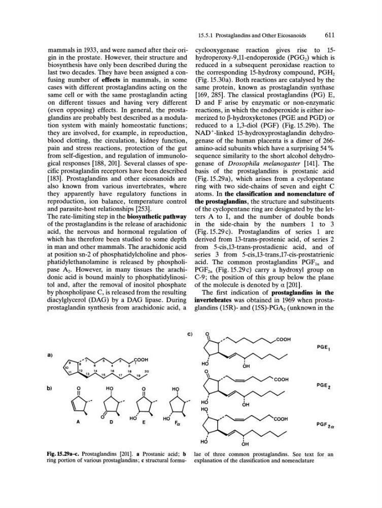
Fig.15.29a-c. Prostaglandins [201]. a Prostanic acid; b
ring portion of various prostaglandins; c structural formu15.5.1 Prostaglandins and Other Eicosanoids
611
cyclooxygenase reaction gives rise to 15hydroperoxy-9,1l-endoperoxide (PGGz) which is
reduced in a subsequent peroxidase reaction to
the corresponding IS-hydroxy compound, PGHz
(Fig. 15.30a). Both reactions are catalysed by the
same protein, known as prostaglandin synthase
[169, 285]. The classical prostaglandins (PG) E,
D and F arise by enzymatic or non-enzymatic
reactions, in which the endoperoxide is either isomerized to ~-hydroxyketones (PGE and PGD) or
reduced to a 1,3-diol (PGF) (Fig. 15.29b). The
NAD+ -linked 15-hydroxyprostaglandin dehydrogenase of the human placenta is a dimer of 266amino-acid subunits which have a surprising 54 %
sequence similarity to the short alcohol dehydrogenase of Drosophila melanogaster [141]. The
basis of the prostaglandins is prostanic acid
(Fig. 15.29a), which arises from a cyclopentane
ring with two side-chains of seven and eight C
atoms. In the classification and nomenclature of
the prostaglandins, the structure and substituents
of the cyclopentane ring are designated by the letters A to I, and the number of double bonds
in the side-chain by the numbers 1 to 3
(Fig. 15.29 c). Prostaglandins of series 1 are
derived from 13-trans-prostenic acid, of series 2
from 5-cis,13-trans-prostadienic acid, and of
series 3 from 5-cis,13-trans,17-cis-prostatrienic
acid. The common prostaglandins PGF1a and
PGFza (Fig. 15.29 c) carry a hydroxyl group on
C-9; the position of this group below the plane
of the molecule is denoted by a [201].
The first indication of prostaglandins in the
invertebrates was obtained in 1969 when prostaglandins (15R)- and (15S)-PGAz (unknown in the
HO
\
\
,'~COOH
.
OH
"~COOH
.
I
OH
PGF 2a
lae of three common prostaglandins. See text for an
explanation of the classification and nomenclature