

is present in vertebrate brain with the highest
concentrations occurring in the amphibians.
Carnosine is synthesized by carnosine synthetase; this forms an enzymes-l3-alanyladenyl complex with the consumption of ATP, and this then
reacts with histidine. The highest enzyme activities are found in the rat, mouse and chicken in
the skeletal muscle and heart, and in the leopard
frog Rana pipiens in the liver. Carnosine can be
methylated to anserine by an S-adenosylmethionine: carnosine methyltransferase which,
however, is not found in all vertebrates. The histidine dipeptides are cleaved by dipeptidases in the
kidney, liver, muscle, brain and other organs. Porcine kidney contains a homocarnosinase with a
broad specificity and a carnosinase with a narrower specificity. The so-called anserinase and Nacetylhistidinase from fish muscle and brain
resemble porcine homocarnosinase in their broad
specificity [1].
12.11 Purine and Pyrimidine Compounds
Every cell contains a complex spectrum of low
molecular weight purine and pyrimidine compounds, which are components or degradation
products of nucleic acids; they function variously
as coenzymes or cosubstrates, secondary messengers, and nitrogenous waste products. Little
variation is encountered and there is usually no
explanation of its biological significance. The
nucleosides with unusual structures, found in several sponges and sea snails, are dealt with as
products of secondary metabolism in Chapter 19.
12.11.1 Biosynthesis of Purine Nucleotides
447
U.ll.1 Biosynthesis of Purine Nucleotides
Purine nucleotides are synthesized either de novo
or from preformed purine bases or nucleosides.
De novo synthesis is not possible in many animals, in which case purine compounds are essential nutrients. In other cases, both synthetic pathways are made use of and their relative importance is usually difficult to estimate. The primary
product of de novo purine synthesis is IMP. The
origins of the individual atoms of the purine ring
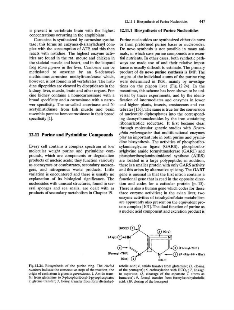
were determined in 1956, mainly by investigations on the pigeon liver (Fig. 12.24). In the
meantime, this scheme has been shown to be universal by tracer experiments, and by the identification of intermediates and enzymes in lower
and higher plants, insects, crustaceans and vertebrates [156]. The same is true for the conversion
of nucleotide diphosphates into the corresponding deoxyribonucleotides by the iron-containing
ribonucleotide reductase. It first became clear
through molecular genetic studies with Drosophila melanogaster that multifunctional enzymes
play an important role in both purine and pyrimidine biosynthesis. The activities of phosphoribosylamineglycine ligase (GARS), phosphoribosylglycine amide formyltransferase (GART) and
phosphoribosylaminoimidazol synthase (AIRS)
are located in a large polypeptide; in addition,
there is a smaller protein with only GARS activity
and this arises by alternative splicing. The GART
gene is unusual in that the first intron contains a
functional gene that is read in the opposite direction and codes for a cuticular protein (p. 15).
There is also a human gene which codes for these
three enzyme activities; in the avian liver, two
enzyme activities of tetrahydrofolate metabolism
are apparently also present on the equivalent protein complex [107]. The dual function of purine as
a nucleic acid component and excretion product is
(HC03) 0 ... 5?
2 (Gly)
(Asp)(D-HN
0-~
i j-::-":"'::-.=NJ
U ).0 (fo,myl-THf)
(Formyl-THF)
N
~
(Gin) CD
N -CD (P-Rib-PP +Gln)
I
Fig. U.24. Biosynthesis of the purine ring. The circled
numbers indicate the consecutive steps of the reaction; the
origin of each atom is given in parentheses. 1, Amido transfer from glutamine to 5-phosphoribosyl-1-pyrophosphate;
2, glycine transfer; 3, formyl transfer from formyltetrahydRib-P
rofolic acid; 4, amido transfer from glutamine; (5, closing
of the pentagon); 6, carboxylation with HC03-; 7, linkage
to aspartate; (8, cleavage of the aspartate C atoms as
fumarate); 9, formyl transfer from formyltetrahydrofolic
acid; (10, closing of the hexagon)