

12.9 Phosphagens and Other Guanidine Compounds
439
COOH
I
CH 3 COOH
I
I
Ornithine
H2N-CCCC-NH2
Adenosine -S-CCC-NH2
0j
CO2 --~
@J
C02--~ TH3
Putrescine
H2N-CCCC-NH2
Adenosine -Sfr-c-cc-r-N-H-'21
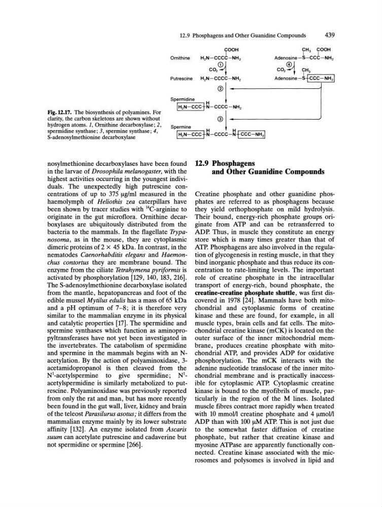
Fig.l2.17. The biosynthesis of polyamines. For
clarity, the carbon skeletons are shown without
hydrogen atoms. 1, Ornithine decarboxylase; 2,
spermidine synthase; 3, spermine synthase; 4,
S-adenosylmethionine decarboxylase
@I :
Spermidine
hN-CCC+~-C:C-NH'
s,,~;"'
j
I H2N-CCCt~-cccc-~fCCC-NH21
nosylmethionine decarboxylases have been found
in the larvae of Drosophila melanogaster, with the
highest activities occurring in the youngest individuals. The unexpectedly high putrescine concentrations of up to 375 !-tg/ml measured in the
haemolymph of Heliothis zea caterpillars have
been shown by tracer studies with 14C-arginine to
originate in the gut microflora. Ornithine decarboxylases are ubiquitously distributed from the
bacteria to the mammals. In the flagellate Trypanosoma, as in the mouse, they are cytoplasmic
dimeric proteins of 2 x 45 kDa. In contrast, in the
nematodes Caenorhabditis elegans and Haemonchus contortus they are membrane bound. The
enzyme from the ciliate Tetrahymena pyriformis is
activated by phosphorylation [129, 140, 183, 216].
The S-adenosylmethionine decarboxylase isolated
from the mantle, hepatopancreas and foot of the
edible mussel Mytilus edulis has a mass of 65 kDa
and a pH optimum of 7-8; it is therefore very
similar to the mammalian enzyme in its physical
and catalytic properties [17]. The spermidine and
spermine synthases which function as aminopropyltransferases have not yet been investigated in
the invertebrates. The catabolism of spermidine
and spermine in the mammals begins with an Nacetylation. By the action of polyaminoxidase, 3acetamidopropanol is then cleaved from the
Nl-acetylspermine to give spermidine; N 1 _
acetylspermidine is similarly metabolized to putrescine. Polyaminoxidase was previously reported
from only the rat and man, but has more recently
been found in the gut wall, liver, kidney and brain
of the teleost Parasilurus asotus; it differs from the
mammalian enzyme mainly by its lower substrate
affinity [132]. An enzyme isolated from Ascaris
suum can acetylate putrescine and cadaverine but
not spermidine or spermine [266].
U.9 Phosphagens
and Other Guanidine Compounds
Creatine phosphate and other guanidine phosphates are referred to as phosphagens because
they yield orthophosphate on mild hydrolysis.
Their bound, energy-rich phosphate groups originate from ATP and can be retransferred to
ADP. Thus, in muscle they constitute an energy
store which is many times greater than that of
ATP. Phosphagens are also involved in the regulation of glycogenesis in resting muscle, in that they
bind inorganic phosphate and thus reduce its concentration to rate-limiting levels. The important
role of creatine phosphate in the intracellular
transport of energy-rich, bound phosphate, the
creatine-creatine phosphate shuttle, was first discovered in 1978 [24]. Mammals have both mito~hondrial and cytoplasmic forms of creatine
kinase and these are found, for example, in all
muscle types, brain cells and fat cells. The mitochondrial creatine kinase (mCK) is located on the
outer surface of the inner mitochondrial membrane, produces creatine phosphate with mitochondrial ATP, and provides ADP for oxidative
phosphorylation. The mCK interacts with the
adenine nucleotide translocase of the inner mitochondrial membrane and is practically inaccessible for cytoplasmic ATP. Cytoplasmic creatine
kinase is bound to the myofibrils of muscle, particularly in the region of the M lines. Isolated
muscle fibres contract more rapidly when treated
with 10 mmolll creatine phosphate and 4 !-tmolll
ADP than with 100 !-tM ATP. This is not just due
to the somewhat faster diffusion of creatine
phosphate, but rather that creatine kinase and
myosine ATPase are apparently functionally connected. Creatine kinase associated with the microsomes and polysomes is involved in lipid and
439
COOH
I
CH 3 COOH
I
I
Ornithine
H2N-CCCC-NH2
Adenosine -S-CCC-NH2
0j
CO2 --~
@J
C02--~ TH3
Putrescine
H2N-CCCC-NH2
Adenosine -Sfr-c-cc-r-N-H-'21
Fig.l2.17. The biosynthesis of polyamines. For
clarity, the carbon skeletons are shown without
hydrogen atoms. 1, Ornithine decarboxylase; 2,
spermidine synthase; 3, spermine synthase; 4,
S-adenosylmethionine decarboxylase
@I :
Spermidine
hN-CCC+~-C:C-NH'
s,,~;"'
j
I H2N-CCCt~-cccc-~fCCC-NH21
nosylmethionine decarboxylases have been found
in the larvae of Drosophila melanogaster, with the
highest activities occurring in the youngest individuals. The unexpectedly high putrescine concentrations of up to 375 !-tg/ml measured in the
haemolymph of Heliothis zea caterpillars have
been shown by tracer studies with 14C-arginine to
originate in the gut microflora. Ornithine decarboxylases are ubiquitously distributed from the
bacteria to the mammals. In the flagellate Trypanosoma, as in the mouse, they are cytoplasmic
dimeric proteins of 2 x 45 kDa. In contrast, in the
nematodes Caenorhabditis elegans and Haemonchus contortus they are membrane bound. The
enzyme from the ciliate Tetrahymena pyriformis is
activated by phosphorylation [129, 140, 183, 216].
The S-adenosylmethionine decarboxylase isolated
from the mantle, hepatopancreas and foot of the
edible mussel Mytilus edulis has a mass of 65 kDa
and a pH optimum of 7-8; it is therefore very
similar to the mammalian enzyme in its physical
and catalytic properties [17]. The spermidine and
spermine synthases which function as aminopropyltransferases have not yet been investigated in
the invertebrates. The catabolism of spermidine
and spermine in the mammals begins with an Nacetylation. By the action of polyaminoxidase, 3acetamidopropanol is then cleaved from the
Nl-acetylspermine to give spermidine; N 1 _
acetylspermidine is similarly metabolized to putrescine. Polyaminoxidase was previously reported
from only the rat and man, but has more recently
been found in the gut wall, liver, kidney and brain
of the teleost Parasilurus asotus; it differs from the
mammalian enzyme mainly by its lower substrate
affinity [132]. An enzyme isolated from Ascaris
suum can acetylate putrescine and cadaverine but
not spermidine or spermine [266].
U.9 Phosphagens
and Other Guanidine Compounds
Creatine phosphate and other guanidine phosphates are referred to as phosphagens because
they yield orthophosphate on mild hydrolysis.
Their bound, energy-rich phosphate groups originate from ATP and can be retransferred to
ADP. Thus, in muscle they constitute an energy
store which is many times greater than that of
ATP. Phosphagens are also involved in the regulation of glycogenesis in resting muscle, in that they
bind inorganic phosphate and thus reduce its concentration to rate-limiting levels. The important
role of creatine phosphate in the intracellular
transport of energy-rich, bound phosphate, the
creatine-creatine phosphate shuttle, was first discovered in 1978 [24]. Mammals have both mito~hondrial and cytoplasmic forms of creatine
kinase and these are found, for example, in all
muscle types, brain cells and fat cells. The mitochondrial creatine kinase (mCK) is located on the
outer surface of the inner mitochondrial membrane, produces creatine phosphate with mitochondrial ATP, and provides ADP for oxidative
phosphorylation. The mCK interacts with the
adenine nucleotide translocase of the inner mitochondrial membrane and is practically inaccessible for cytoplasmic ATP. Cytoplasmic creatine
kinase is bound to the myofibrils of muscle, particularly in the region of the M lines. Isolated
muscle fibres contract more rapidly when treated
with 10 mmolll creatine phosphate and 4 !-tmolll
ADP than with 100 !-tM ATP. This is not just due
to the somewhat faster diffusion of creatine
phosphate, but rather that creatine kinase and
myosine ATPase are apparently functionally connected. Creatine kinase associated with the microsomes and polysomes is involved in lipid and