

396
11 Extracellular Structural and Secretory Proteins
amino acid composition quite different from that
of the large subunit [Table 11.4 (1b)]. The light
fibroin chain, encoded by its own gene, is 244
amino acids in length. This includes three cysteine
residues, of which two form an internal disulphide bridge, and the third is responsible for the
link to the heavy chain [142]. Fibroin contains an
asparagine-linked oligosaccharide of N-acetylglucosamine and mannose. A gene for the large
fibroin chain, sequenced from the Japanese oak
eggar Antheraea yamamai, consists of two exons,
as in Bombyx mori. The two genes show close
agreement in a region covering 300 bp of the 5'NT region and the 70 bp of the first small exon,
but do not show close agreement in the second
exon, which encodes the major part of the fibroin
sequence. Thus, the 5'-NTregion is important for
expression of the fibroin gene in the posterior silk
gland, and is highly conserved [126].
Much less is known about sericin, the adhesive
protein of the silk thread. Two-thirds of the amino
acids are serine, glycine and aspartic acid [Table 11.4 (1 c)]; in addition to N-glycosidic asparagine-bound carbohydrate, as found in fibroin,
sericin also contains O-glycosidic serine- or
threonine-bound oligo saccharides of N-acetylgalactosamine and galactose. Six sericin polypeptides with molecular masses between 65 and
400 kDa have been isolated from the silk glands of
Bombyx mori; these are the result of alternative
splicing of transcripts from only two genes [80].
The amino acid compositions, conformations
and mechanical properties of the various silks are
extraordinarily varied. The amino acid spectrum
does not always correspond to the scheme in
Bombyx mori, where glycine, alanine and serine
predominate (Table 11.4). Thus, there are silks
high in glutamine, as in those of Digelansinus
diversiipes and Arge ustalata [114], and the silk of
the lepidopteran Calpodes ethlius contains no less
than 25.5 % proline. The fibroins can be ordered
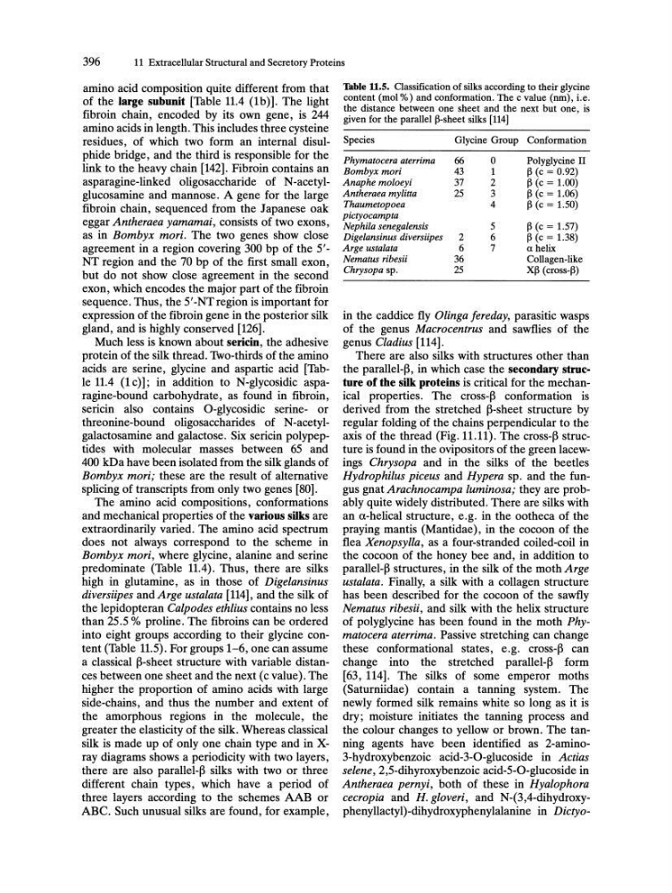
into eight groups according to their glycine content (Table 11.5). For groups 1-6, one can assume
a classical ~-sheet structure with variable distances between one sheet and the next (c value). The
higher the proportion of amino acids with large
side-chains, and thus the number and extent of
the amorphous regions in the molecule, the
greater the elasticity of the silk. Whereas classical
silk is made up of only one chain type and in Xray diagrams shows a periodicity with two layers,
there are also parallel-~ silks with two or three
different chain types, which have a period of
three layers according to the schemes AAB or
ABC. Such unusual silks are found, for example,
Table 11.5. Classification of silks according to their glycine
content (mol %) and conformation. The c value (nm), i.e.
the distance between one sheet and the next but one, is
given for the parallel ~-sheet silks [114]
Species
Glycine Group Conformation
Phymatocera aterrima
66
0
Polyglycine II
Bombyx mori
43
1
~ (c = 0.92)
Anaphe moloeyi
37
2
~ (c = 1.00)
Antheraea mylitta
25
3
~ (c = 1.06)
Thaumetopoea
4
~ (c = 1.50)
pictyocampta
Nephila senegalensis
5
~ (c = 1.57)
Digelansinus diversiipes
2
6
~ (c = 1.38)
Arge ustalata
6
7
a helix
Nematus ribesii
36
Collagen-like
Chrysopa sp.
25
X~ (cross-~)
in the caddice fly Olinga fereday, parasitic wasps
of the genus Macrocentrus and sawflies of the
genus Cladius [114].
There are also silks with structures other than
the parallel-~, in which case the secondary structure of the silk proteins is critical for the mechanical properties. The cross-~ conformation is
derived from the stretched ~-sheet structure by
regular folding of the chains perpendicular to the
axis of the thread (Fig. 11.11). The cross-~ structure is found in the ovipositors of the green lacewings Chrysopa and in the silks of the beetles
Hydrophilus piceus and Hypera sp. and the fungus gnat Arachnocampa luminosa; they are probably quite widely distributed. There are silks with
an a-helical structure, e.g. in the ootheca of the
praying mantis (Mantidae), in the cocoon of the
flea Xenopsylla, as a four-stranded coiled-coil in
the cocoon of the honey bee and, in addition to
parallel-~ structures, in the silk of the moth Arge
ustalata. Finally, a silk with a collagen structure
has been described for the cocoon of the sawfly
Nematus ribesii, and silk with the helix structure
of poly glycine has been found in the moth Phymatocera aterrima. Passive stretching can change
these conformational states, e.g. cross-~ can
change into the stretched parallel-~ form
[63, 114]. The silks of some emperor moths
(Saturniidae) contain a tanning system. The
newly formed silk remains white so long as it is
dry; moisture initiates the tanning process and
the colour changes to yellow or brown. The tanning agents have been identified as 2-amino3-hydroxybenzoic acid-3-0-glucoside in Actias
selene, 2,5-dihyroxybenzoic acid-5-0-glucoside in
Antheraea pernyi, both of these in Hyalophora
cecropia and H. gloveri, and N-(3,4-dihydroxyphenyllactyl)-dihydroxyphenylalanine in Dictyo-
11 Extracellular Structural and Secretory Proteins
amino acid composition quite different from that
of the large subunit [Table 11.4 (1b)]. The light
fibroin chain, encoded by its own gene, is 244
amino acids in length. This includes three cysteine
residues, of which two form an internal disulphide bridge, and the third is responsible for the
link to the heavy chain [142]. Fibroin contains an
asparagine-linked oligosaccharide of N-acetylglucosamine and mannose. A gene for the large
fibroin chain, sequenced from the Japanese oak
eggar Antheraea yamamai, consists of two exons,
as in Bombyx mori. The two genes show close
agreement in a region covering 300 bp of the 5'NT region and the 70 bp of the first small exon,
but do not show close agreement in the second
exon, which encodes the major part of the fibroin
sequence. Thus, the 5'-NTregion is important for
expression of the fibroin gene in the posterior silk
gland, and is highly conserved [126].
Much less is known about sericin, the adhesive
protein of the silk thread. Two-thirds of the amino
acids are serine, glycine and aspartic acid [Table 11.4 (1 c)]; in addition to N-glycosidic asparagine-bound carbohydrate, as found in fibroin,
sericin also contains O-glycosidic serine- or
threonine-bound oligo saccharides of N-acetylgalactosamine and galactose. Six sericin polypeptides with molecular masses between 65 and
400 kDa have been isolated from the silk glands of
Bombyx mori; these are the result of alternative
splicing of transcripts from only two genes [80].
The amino acid compositions, conformations
and mechanical properties of the various silks are
extraordinarily varied. The amino acid spectrum
does not always correspond to the scheme in
Bombyx mori, where glycine, alanine and serine
predominate (Table 11.4). Thus, there are silks
high in glutamine, as in those of Digelansinus
diversiipes and Arge ustalata [114], and the silk of
the lepidopteran Calpodes ethlius contains no less
than 25.5 % proline. The fibroins can be ordered
into eight groups according to their glycine content (Table 11.5). For groups 1-6, one can assume
a classical ~-sheet structure with variable distances between one sheet and the next (c value). The
higher the proportion of amino acids with large
side-chains, and thus the number and extent of
the amorphous regions in the molecule, the
greater the elasticity of the silk. Whereas classical
silk is made up of only one chain type and in Xray diagrams shows a periodicity with two layers,
there are also parallel-~ silks with two or three
different chain types, which have a period of
three layers according to the schemes AAB or
ABC. Such unusual silks are found, for example,
Table 11.5. Classification of silks according to their glycine
content (mol %) and conformation. The c value (nm), i.e.
the distance between one sheet and the next but one, is
given for the parallel ~-sheet silks [114]
Species
Glycine Group Conformation
Phymatocera aterrima
66
0
Polyglycine II
Bombyx mori
43
1
~ (c = 0.92)
Anaphe moloeyi
37
2
~ (c = 1.00)
Antheraea mylitta
25
3
~ (c = 1.06)
Thaumetopoea
4
~ (c = 1.50)
pictyocampta
Nephila senegalensis
5
~ (c = 1.57)
Digelansinus diversiipes
2
6
~ (c = 1.38)
Arge ustalata
6
7
a helix
Nematus ribesii
36
Collagen-like
Chrysopa sp.
25
X~ (cross-~)
in the caddice fly Olinga fereday, parasitic wasps
of the genus Macrocentrus and sawflies of the
genus Cladius [114].
There are also silks with structures other than
the parallel-~, in which case the secondary structure of the silk proteins is critical for the mechanical properties. The cross-~ conformation is
derived from the stretched ~-sheet structure by
regular folding of the chains perpendicular to the
axis of the thread (Fig. 11.11). The cross-~ structure is found in the ovipositors of the green lacewings Chrysopa and in the silks of the beetles
Hydrophilus piceus and Hypera sp. and the fungus gnat Arachnocampa luminosa; they are probably quite widely distributed. There are silks with
an a-helical structure, e.g. in the ootheca of the
praying mantis (Mantidae), in the cocoon of the
flea Xenopsylla, as a four-stranded coiled-coil in
the cocoon of the honey bee and, in addition to
parallel-~ structures, in the silk of the moth Arge
ustalata. Finally, a silk with a collagen structure
has been described for the cocoon of the sawfly
Nematus ribesii, and silk with the helix structure
of poly glycine has been found in the moth Phymatocera aterrima. Passive stretching can change
these conformational states, e.g. cross-~ can
change into the stretched parallel-~ form
[63, 114]. The silks of some emperor moths
(Saturniidae) contain a tanning system. The
newly formed silk remains white so long as it is
dry; moisture initiates the tanning process and
the colour changes to yellow or brown. The tanning agents have been identified as 2-amino3-hydroxybenzoic acid-3-0-glucoside in Actias
selene, 2,5-dihyroxybenzoic acid-5-0-glucoside in
Antheraea pernyi, both of these in Hyalophora
cecropia and H. gloveri, and N-(3,4-dihydroxyphenyllactyl)-dihydroxyphenylalanine in Dictyo-