

v
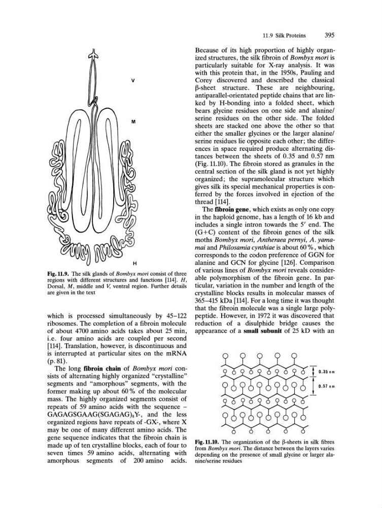
Fig.ll.9. The silk glands of Bombyx mori consist of three
regions with different structures and functions [114]. H,
Dorsal, M, middle and V, ventral region. Further details
are given in the text
which is processed simultaneously by 45-122
ribosomes. The completion of a fibroin molecule
of about 4700 amino acids takes about 25 min,
i.e. four amino acids are coupled per second
[114]. Translation, however, is discontinuous and
is interrupted at particular sites on the mRNA
(p.81).
The long fibroin chain of Bombyx mori consists of alternating highly organized "crystalline"
segments and "amorphous" segments, with the
former making up about 60 % of the molecular
mass. The highly organized segments consist of
repeats of 59 amino acids with the sequence -
GAGAGSGAAG(SGAGAG)8Y-, and the less
organized regions have repeats of -GX-, where X
may be one of many different amino acids. The
gene sequence indicates that the fibroin chain is
made up of ten crystalline blocks, each of four to
seven times 59 amino acids, alternating with
amorphous segments of 200 amino acids.
11.9 Silk Proteins
395
Because of its high proportion of highly organized structures, the silk fibroin of Bombyx mori is
particularly suitable for X-ray analysis. It was
with this protein that, in the 1950s, Pauling and
Corey discovered and described the classical
B-sheet structure. These are neighbouring,
antiparallel-orientated peptide chains that are linked by H-bonding into a folded sheet, which
bears glycine residues on one side and alanine!
serine residues on the other side. The folded
sheets are stacked one above the other so that
either the smaller glycines or the larger alanine!
serine residues lie opposite each other; the differences in space required produce alternating distances between the sheets of 0.35 and 0.57 nm
(Fig. 11.10). The fibroin stored as granules in the
central section of the silk gland is not yet highly
organized; the supramolecular structure which
gives silk its special mechanical properties is conferred by the forces involved in ejection of the
thread [114].
The fibroin gene, which exists as only one copy
in the haploid genome, has a length of 16 kb and
includes a single intron towards the 5' end. The
(G+C) content of the fibroin genes of the silk
moths Bombyx mori, Antheraea pernyi, A. yamamai and Philosamia cynthiae is about 60 % , which
corresponds to the codon preference of GGN for
alanine and GCN for glycine [126]. Comparison
of various lines of Bombyx mori reveals considerable polymorphism of the fibroin gene. In particular, variation in the number and length of the
crystalline blocks results in molecular masses of
365-415 kDa [114]. For a long time it was thought
that the fibroin molecule was a single large polypeptide. However, in 1972 it was discovered that
reduction of a disulphide bridge causes the
appearance of a small subunit of 25 kD with an
T 0.35 nm
t 0.57nm
Fig.ll.l0. The organization of the ~-sheets in silk fibres
from Bombyx mori. The distance between the layers varies
depending on the presence of small glycine or larger alanine/serine residues
Fig.ll.9. The silk glands of Bombyx mori consist of three
regions with different structures and functions [114]. H,
Dorsal, M, middle and V, ventral region. Further details
are given in the text
which is processed simultaneously by 45-122
ribosomes. The completion of a fibroin molecule
of about 4700 amino acids takes about 25 min,
i.e. four amino acids are coupled per second
[114]. Translation, however, is discontinuous and
is interrupted at particular sites on the mRNA
(p.81).
The long fibroin chain of Bombyx mori consists of alternating highly organized "crystalline"
segments and "amorphous" segments, with the
former making up about 60 % of the molecular
mass. The highly organized segments consist of
repeats of 59 amino acids with the sequence -
GAGAGSGAAG(SGAGAG)8Y-, and the less
organized regions have repeats of -GX-, where X
may be one of many different amino acids. The
gene sequence indicates that the fibroin chain is
made up of ten crystalline blocks, each of four to
seven times 59 amino acids, alternating with
amorphous segments of 200 amino acids.
11.9 Silk Proteins
395
Because of its high proportion of highly organized structures, the silk fibroin of Bombyx mori is
particularly suitable for X-ray analysis. It was
with this protein that, in the 1950s, Pauling and
Corey discovered and described the classical
B-sheet structure. These are neighbouring,
antiparallel-orientated peptide chains that are linked by H-bonding into a folded sheet, which
bears glycine residues on one side and alanine!
serine residues on the other side. The folded
sheets are stacked one above the other so that
either the smaller glycines or the larger alanine!
serine residues lie opposite each other; the differences in space required produce alternating distances between the sheets of 0.35 and 0.57 nm
(Fig. 11.10). The fibroin stored as granules in the
central section of the silk gland is not yet highly
organized; the supramolecular structure which
gives silk its special mechanical properties is conferred by the forces involved in ejection of the
thread [114].
The fibroin gene, which exists as only one copy
in the haploid genome, has a length of 16 kb and
includes a single intron towards the 5' end. The
(G+C) content of the fibroin genes of the silk
moths Bombyx mori, Antheraea pernyi, A. yamamai and Philosamia cynthiae is about 60 % , which
corresponds to the codon preference of GGN for
alanine and GCN for glycine [126]. Comparison
of various lines of Bombyx mori reveals considerable polymorphism of the fibroin gene. In particular, variation in the number and length of the
crystalline blocks results in molecular masses of
365-415 kDa [114]. For a long time it was thought
that the fibroin molecule was a single large polypeptide. However, in 1972 it was discovered that
reduction of a disulphide bridge causes the
appearance of a small subunit of 25 kD with an
T 0.35 nm
t 0.57nm
Fig.ll.l0. The organization of the ~-sheets in silk fibres
from Bombyx mori. The distance between the layers varies
depending on the presence of small glycine or larger alanine/serine residues