

380
11 Extracellular Structural and Secretory Proteins
···OC NH···
, /
CH
I
1H Jbj
¥ 6H
CH2
I
CH
/ ,
···HN co···
Bistyrosine
CH 2
I
CH
/ ,
···HN co···
Tertyrosine
CH 2
I
CH
···OC NH···
,/ CH I
CH 2
···HNCO ...
Isotertyrosine
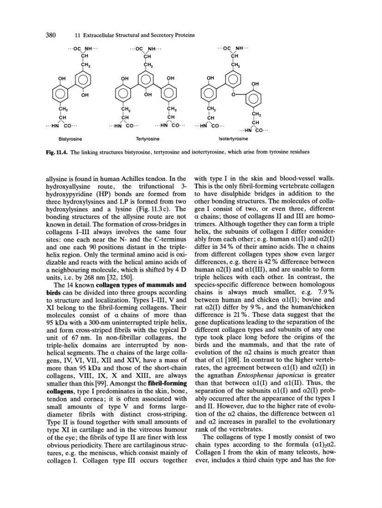
Fig.11.4. The linking structures bistyrosine, tertyrosine and isotertyrosine, which arise from tyrosine residues
allysine is found in human Achilles tendon. In the
hydroxyallysine route, the trifunctional 3hydroxypyridine (HP) bonds are formed from
three hydroxylysines and LP is formed from two
hydroxylysines and a lysine (Fig. 11.3 c). The
bonding structures of the allysine route are not
known in detail. The formation of cross-bridges in
collagens I-III always involves the same four
sites: one each near the N- and the C-terminus
and one each 90 positions distant in the triplehelix region. Only the terminal amino acid is oxidizable and reacts with the helical amino acids of
a neighbouring molecule, which is shifted by 4 D
units, i.e. by 268 nm [32, 150].
The 14 known collagen types of mammals and
birds can be divided into three groups according
to structure and localization. Types I-III, V and
XI belong to the fibril-forming collagens. Their
molecules consist of a chains of more than
95 kDa with a 300-nm uninterrupted triple helix,
and form cross-striped fibrils with the typical D
unit of 67 nm. In non-fibrillar collagens, the
triple-helix domains are interrupted by nonhelical segments. The a chains of the large collagens, IV, VI, VII, XII and XlV, have a mass of
more than 95 kDa and those of the short-chain
collagens, VIII, IX, X and XIII, are always
smaller than this [99]. Amongst the fibril.forming
collagens, type I predominates in the skin, bone,
tendon and cornea; it is often associated with
small amounts of type V and forms largediameter fibrils with distinct cross-striping.
Type II is found together with small amounts of
type XI in cartilage and in the vitreous humour
of the eye; the fibrils of type II are finer with less
obvious periodicity. There are cartilaginous structures, e.g. the meniscus, which consist mainly of
collagen I. Collagen type III occurs together
with type I in the skin and blood-vessel walls.
This is the only fibril-forming vertebrate collagen
to have disulphide bridges in addition to the
other bonding structures. The molecules of collagen I consist of two, or even three, different
a chains; those of collagens II and III are homotrimers. Although together they can form a triple
helix, the subunits of collagen I differ considerably from each other; e.g. human a1(1) and aZ(I)
differ in 34 % of their amino acids. The a chains
from different collagen types show even larger
differences, e.g. there is 42 % difference between
human aZ(I) and a1(1I1) , and are unable to form
triple helices with each other. In contrast, the
species-specific difference between homologous
chains is always much smaller, e.g. 7.9 %
between human and chicken a1(1); bovine and
rat aZ(I) differ by 9 %, and the human/chicken
difference is Z1 %. These data suggest that the
gene duplications leading to the separation of the
different collagen types and subunits of anyone
type took place long before the origins of the
birds and the mammals, and that the rate of
evolution of the aZ chains is much greater than
that of a1 [108]. In contrast to the higher vertebrates, the agreement between a1(1) and aZ(I) in
the agnathan Entosphenus japonicus is greater
than that between a1(I) and a1(1I). Thus, the
separation of the subunits a1(1) and aZ(I) probably occurred after the appearance of the types I
and II. However, due to the higher rate of evolution of the aZ chains, the difference between a1
and aZ increases in parallel to the evolutionary
rank of the vertebrates.
The collagens of type I mostly consist of two
chain types according to the formula (a1)2aZ.
Collagen I from the skin of many teleosts, however, includes a third chain type and has the for-
11 Extracellular Structural and Secretory Proteins
···OC NH···
, /
CH
I
1H Jbj
¥ 6H
CH2
I
CH
/ ,
···HN co···
Bistyrosine
CH 2
I
CH
/ ,
···HN co···
Tertyrosine
CH 2
I
CH
···OC NH···
,/ CH I
CH 2
···HNCO ...
Isotertyrosine
Fig.11.4. The linking structures bistyrosine, tertyrosine and isotertyrosine, which arise from tyrosine residues
allysine is found in human Achilles tendon. In the
hydroxyallysine route, the trifunctional 3hydroxypyridine (HP) bonds are formed from
three hydroxylysines and LP is formed from two
hydroxylysines and a lysine (Fig. 11.3 c). The
bonding structures of the allysine route are not
known in detail. The formation of cross-bridges in
collagens I-III always involves the same four
sites: one each near the N- and the C-terminus
and one each 90 positions distant in the triplehelix region. Only the terminal amino acid is oxidizable and reacts with the helical amino acids of
a neighbouring molecule, which is shifted by 4 D
units, i.e. by 268 nm [32, 150].
The 14 known collagen types of mammals and
birds can be divided into three groups according
to structure and localization. Types I-III, V and
XI belong to the fibril-forming collagens. Their
molecules consist of a chains of more than
95 kDa with a 300-nm uninterrupted triple helix,
and form cross-striped fibrils with the typical D
unit of 67 nm. In non-fibrillar collagens, the
triple-helix domains are interrupted by nonhelical segments. The a chains of the large collagens, IV, VI, VII, XII and XlV, have a mass of
more than 95 kDa and those of the short-chain
collagens, VIII, IX, X and XIII, are always
smaller than this [99]. Amongst the fibril.forming
collagens, type I predominates in the skin, bone,
tendon and cornea; it is often associated with
small amounts of type V and forms largediameter fibrils with distinct cross-striping.
Type II is found together with small amounts of
type XI in cartilage and in the vitreous humour
of the eye; the fibrils of type II are finer with less
obvious periodicity. There are cartilaginous structures, e.g. the meniscus, which consist mainly of
collagen I. Collagen type III occurs together
with type I in the skin and blood-vessel walls.
This is the only fibril-forming vertebrate collagen
to have disulphide bridges in addition to the
other bonding structures. The molecules of collagen I consist of two, or even three, different
a chains; those of collagens II and III are homotrimers. Although together they can form a triple
helix, the subunits of collagen I differ considerably from each other; e.g. human a1(1) and aZ(I)
differ in 34 % of their amino acids. The a chains
from different collagen types show even larger
differences, e.g. there is 42 % difference between
human aZ(I) and a1(1I1) , and are unable to form
triple helices with each other. In contrast, the
species-specific difference between homologous
chains is always much smaller, e.g. 7.9 %
between human and chicken a1(1); bovine and
rat aZ(I) differ by 9 %, and the human/chicken
difference is Z1 %. These data suggest that the
gene duplications leading to the separation of the
different collagen types and subunits of anyone
type took place long before the origins of the
birds and the mammals, and that the rate of
evolution of the aZ chains is much greater than
that of a1 [108]. In contrast to the higher vertebrates, the agreement between a1(1) and aZ(I) in
the agnathan Entosphenus japonicus is greater
than that between a1(I) and a1(1I). Thus, the
separation of the subunits a1(1) and aZ(I) probably occurred after the appearance of the types I
and II. However, due to the higher rate of evolution of the aZ chains, the difference between a1
and aZ increases in parallel to the evolutionary
rank of the vertebrates.
The collagens of type I mostly consist of two
chain types according to the formula (a1)2aZ.
Collagen I from the skin of many teleosts, however, includes a third chain type and has the for-