

Table 6.1. Human immunoglobulins [23]
IgG
IgGl
IgG2
IgG3
Serum concentration (mg/ml)
5
3
0.5
Quaternary structure
mono mono mono
Size (kDa)
105
150
160
H chain
- designation
yl
l
l
- size (kDa) inc!. carbohydrate
51
51
56
- number of domains
4
4
4
- amino acids in hinge region
15
12
62
mono, monomer; pent, pentamer; di, dimer
tes of different amount and composition. Each
chain consists of a variable (V) region, where the
specific interaction with the antigen occurs, and a
constant (C) region, which is responsible for
effector functions such as binding to the cell surface or complement activation. The chains are
subdivided into domains of about 110 amino
acids, corresponding in their spatial structure to
the Ig homology units mentioned above. The suba)
6.2.1 Basic Structure ofImmunoglobulins
223
IgM
IgA
IgD
IgE
IgG4
IgA1
IgA2
sigA
0.5
1.5
3
0.5
0 . 05
0.03 5. 10- 5
mono pent
mono mono di
mono mono
150
970
160
160
415
175
190
l
f..t
a 1
a 2
a 1 /a 2
[,
E
51
72
57
57
63
72
4
5
4
4
4
5
12
20
7
64
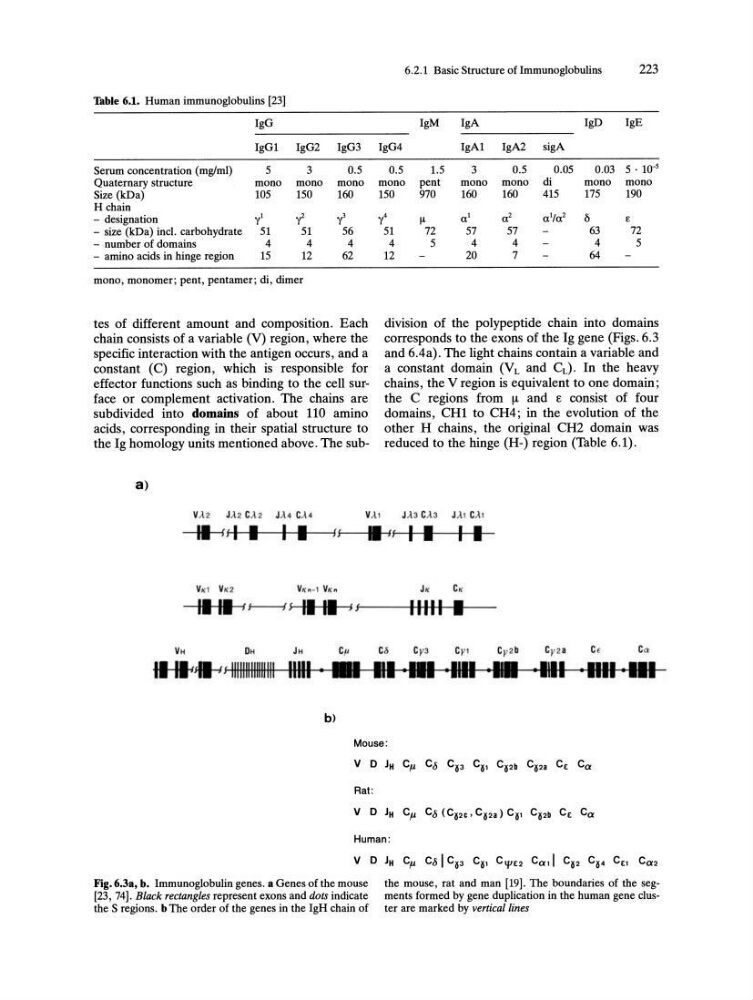
division of the polypeptide chain into domains
corresponds to the exons of the Ig gene (Figs. 6.3
and 6.4a) . The light chains contain a variable and
a constant domain (V Land CL). In the heavy
chains, the V region is equivalent to one domain;
the C regions from !1. and £ consist of four
domains, CHI to CH4; in the evolution of the
other H chains, the original CH2 domain was
reduced to the hinge (H-) region (Table 6.1) .
VA'
II ff I • I I Hf-----i .---';- / f I I I I
II I. 1 J---I f II II JJf----HIII+IIIH-I-I • ...--b)
Mouse :
Rat:
V D JH C il C8 (C~2C • C~2a) C~, C~2b C E Ca
Human :
Fig. 6.3a, b. Immunoglobulin genes. a Genes of the mouse
[23, 74] . Black rectangles represent exons and dots indicate
the S regions. b The order of the genes in the IgH chain of
the mouse, rat and man [19]. The boundaries of the segments formed by gene duplication in the human gene cluster are marked by vertical lines
IgG
IgGl
IgG2
IgG3
Serum concentration (mg/ml)
5
3
0.5
Quaternary structure
mono mono mono
Size (kDa)
105
150
160
H chain
- designation
yl
l
l
- size (kDa) inc!. carbohydrate
51
51
56
- number of domains
4
4
4
- amino acids in hinge region
15
12
62
mono, monomer; pent, pentamer; di, dimer
tes of different amount and composition. Each
chain consists of a variable (V) region, where the
specific interaction with the antigen occurs, and a
constant (C) region, which is responsible for
effector functions such as binding to the cell surface or complement activation. The chains are
subdivided into domains of about 110 amino
acids, corresponding in their spatial structure to
the Ig homology units mentioned above. The suba)
6.2.1 Basic Structure ofImmunoglobulins
223
IgM
IgA
IgD
IgE
IgG4
IgA1
IgA2
sigA
0.5
1.5
3
0.5
0 . 05
0.03 5. 10- 5
mono pent
mono mono di
mono mono
150
970
160
160
415
175
190
l
f..t
a 1
a 2
a 1 /a 2
[,
E
51
72
57
57
63
72
4
5
4
4
4
5
12
20
7
64
division of the polypeptide chain into domains
corresponds to the exons of the Ig gene (Figs. 6.3
and 6.4a) . The light chains contain a variable and
a constant domain (V Land CL). In the heavy
chains, the V region is equivalent to one domain;
the C regions from !1. and £ consist of four
domains, CHI to CH4; in the evolution of the
other H chains, the original CH2 domain was
reduced to the hinge (H-) region (Table 6.1) .
VA'
II ff I • I I Hf-----i .---';- / f I I I I
II I. 1 J---I f II II JJf----HIII+IIIH-I-I • ...--b)
Mouse :
Rat:
V D JH C il C8 (C~2C • C~2a) C~, C~2b C E Ca
Human :
Fig. 6.3a, b. Immunoglobulin genes. a Genes of the mouse
[23, 74] . Black rectangles represent exons and dots indicate
the S regions. b The order of the genes in the IgH chain of
the mouse, rat and man [19]. The boundaries of the segments formed by gene duplication in the human gene cluster are marked by vertical lines