

17.3.2.3 Solid-Phase Cytometry (SPC)
SPC is not very common but it is a very powerful technique for
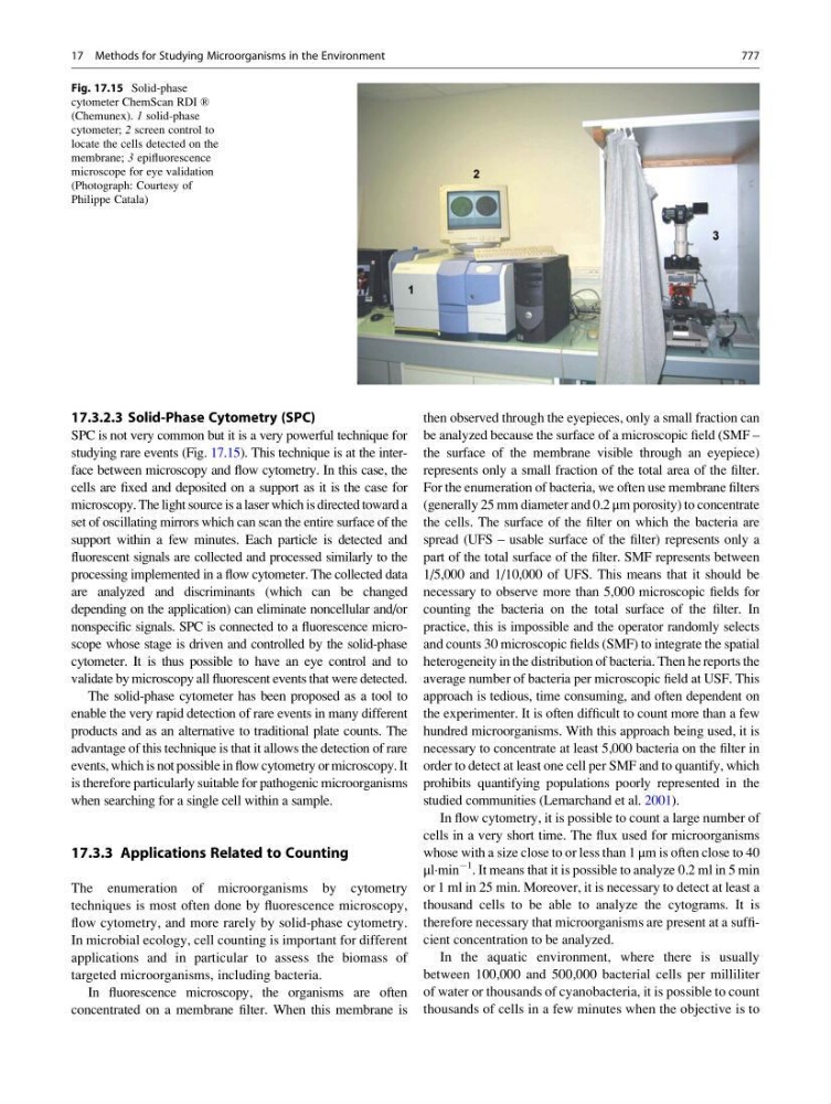
studying rare events (Fig. 17.15). This technique is at the interface between microscopy and flow cytometry. In this case, the
cells are fixed and deposited on a support as it is the case for
microscopy. The light source is a laser which is directed toward a
set of oscillating mirrors which can scan the entire surface of the
support within a few minutes. Each particle is detected and
fluorescent signals are collected and processed similarly to the
processing implemented in a flow cytometer. The collected data
are analyzed and discriminants (which can be changed
depending on the application) can eliminate noncellular and/or
nonspecific signals. SPC is connected to a fluorescence microscope whose stage is driven and controlled by the solid-phase
cytometer. It is thus possible to have an eye control and to
validate by microscopy all fluorescent events that were detected.
The solid-phase cytometer has been proposed as a tool to
enable the very rapid detection of rare events in many different
products and as an alternative to traditional plate counts. The
advantage of this technique is that it allows the detection of rare
events, which is not possible in flow cytometry or microscopy. It
is therefore particularly suitable for pathogenic microorganisms
when searching for a single cell within a sample.
17.3.3 Applications Related to Counting
The enumeration of microorganisms by cytometry
techniques is most often done by fluorescence microscopy,
flow cytometry, and more rarely by solid-phase cytometry.
In microbial ecology, cell counting is important for different
applications and in particular to assess the biomass of
targeted microorganisms, including bacteria.
In fluorescence microscopy, the organisms are often
concentrated on a membrane filter. When this membrane is
then observed through the eyepieces, only a small fraction can
be analyzed because the surface of a microscopic field (SMF –
the surface of the membrane visible through an eyepiece)
represents only a small fraction of the total area of the filter.
For the enumeration of bacteria, we often use membrane filters
(generally 25 mm diameter and 0.2 μm porosity) to concentrate
the cells. The surface of the filter on which the bacteria are
spread (UFS – usable surface of the filter) represents only a
part of the total surface of the filter. SMF represents between
1/5,000 and 1/10,000 of UFS. This means that it should be
necessary to observe more than 5,000 microscopic fields for
counting the bacteria on the total surface of the filter. In
practice, this is impossible and the operator randomly selects
and counts 30 microscopic fields (SMF) to integrate the spatial
heterogeneity in the distribution of bacteria. Then he reports the
average number of bacteria per microscopic field at USF. This
approach is tedious, time consuming, and often dependent on
the experimenter. It is often difficult to count more than a few
hundred microorganisms. With this approach being used, it is
necessary to concentrate at least 5,000 bacteria on the filter in
order to detect at least one cell per SMF and to quantify, which
prohibits quantifying populations poorly represented in the
studied communities (Lemarchand et al. 2001).
In flow cytometry, it is possible to count a large number of
cells in a very short time. The flux used for microorganisms
whose with a size close to or less than 1 μm is often close to 40
μl·min
À1
. It means that it is possible to analyze 0.2 ml in 5 min
or 1 ml in 25 min. Moreover, it is necessary to detect at least a
thousand cells to be able to analyze the cytograms. It is
therefore necessary that microorganisms are present at a sufficient concentration to be analyzed.
In the aquatic environment, where there is usually
between 100,000 and 500,000 bacterial cells per milliliter
of water or thousands of cyanobacteria, it is possible to count
thousands of cells in a few minutes when the objective is to
Fig. 17.15 Solid-phase
cytometer ChemScan RDI ®
(Chemunex). 1 solid-phase
cytometer; 2 screen control to
locate the cells detected on the
membrane; 3 epifluorescence
microscope for eye validation
(Photograph: Courtesy of
Philippe Catala)
17 Methods for Studying Microorganisms in the Environment
777