

obtained with the SEM. In microbial ecology, this technique
is used to describe surfaces, cell morphology, and more
generally the interaction between the cell and its environment. A new generation is derived from SEM microscope: it
is the environmental microscope in which a partial vacuum
is produced at the place of the sample in order to preserve its
“environment”. In this case the metallization is no longer
necessary.
17.3.2.2 Flow Cytometry (FCM)
In cell biology, FCM is a laser-based, biophysical technology employed in cell counting, cell sorting, biomarker
detection, and protein engineering by suspending cells in
a stream of fluid and passing them by an electronic detection apparatus. A beam of light (usually laser light) of a
single wavelength is directed onto a hydrodynamically
focused stream of liquid. A number of detectors are aimed
at the point where the stream passes through the light beam:
one in line with the light beam (Forward Scatter or FSC)
and several perpendicular to it (Side Scatter or SSC) and
one or more fluorescence detectors. Each suspended particle from 0.2 to 150 μm passing through the beam scatters
the ray, and fluorescent chemicals found in the particle or
attached to the particle may be excited into emitting light at
a longer wavelength than the light source. This combination of scattered and fluorescent light is picked up by the
detectors, and, by analyzing fluctuations in brightness at
each detector (one for each fluorescent emission peak), it is
then possible to derive various types of information about
the physical and chemical structure of each individual
particle.
It differs from microscopy because cells are no longer
fixed on a support but driven by a liquid stream, hence the
term “flow.” For a more technical description of the instrument, the reader is referred to one of the many websites
dedicated to flow cytometry platforms.
The measured signals are essentially:
(i) Physical signals which correspond to the properties of
light scattering related to the dimensions of the particle,
its internal structure, or form
(ii) Optical signals which correspond to the constitutive
fluorescence properties for cells that are naturally fluorescent (photosynthetic cells) or whose fluorescence is
induced by a specific staining on the basis of one or
more probes
Each cell is analyzed individually on the basis of several
parameters. Multiparametric data are acquired over
thousands of events per second, and it is thus possible to
obtain statistics on very large populations. These data are
shown as monoparametric histograms or cytograms combining two parameters.
Flow cytometry is a very common technique in microbial
ecology not only for the enumeration of bacteria, pico- and
micro-phytoplankton, and more recently microzooplankton
but also for viruses (even if more difficult). FCM allows
the study and quantification of the properties of cells. This
technique has participated to the development of microbial
ecology by providing informations on the abundance of
microorganisms in a variety of environments and not only to
discover many new groups (Synechococcus, Prochlorococcus,
Ostreococcus, etc.) but also to reveal their importance in
different oceanic regions and in the element cycles in the
sea. Flow cytometers are increasingly onboard oceanographic
vessels and provide near real-time data that are essential to
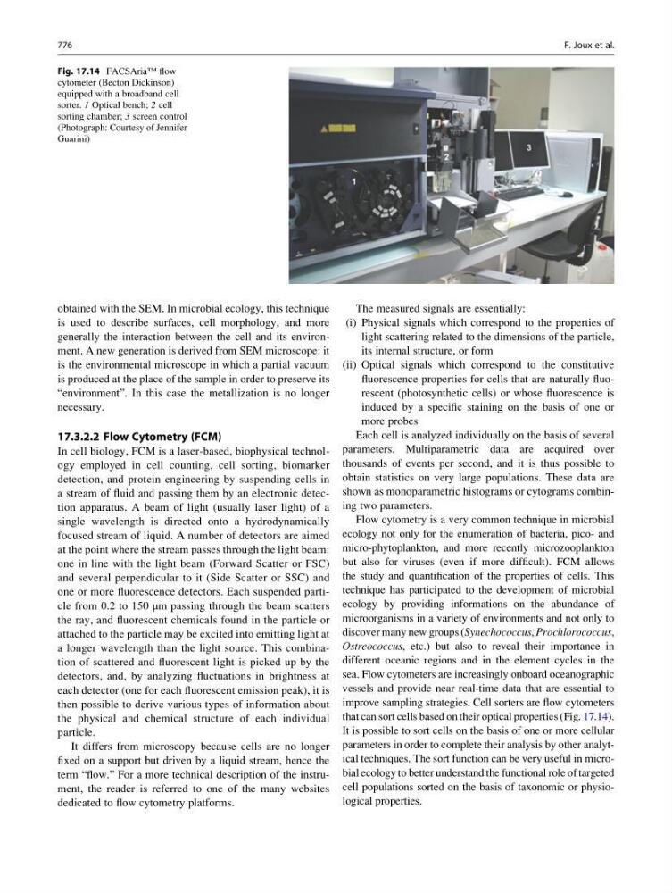
improve sampling strategies. Cell sorters are flow cytometers
that can sort cells based on their optical properties (Fig. 17.14).
It is possible to sort cells on the basis of one or more cellular
parameters in order to complete their analysis by other analytical techniques. The sort function can be very useful in microbial ecology to better understand the functional role of targeted
cell populations sorted on the basis of taxonomic or physiological properties.
Fig. 17.14 FACSAria™ flow
cytometer (Becton Dickinson)
equipped with a broadband cell
sorter. 1 Optical bench; 2 cell
sorting chamber; 3 screen control
(Photograph: Courtesy of Jennifer
Guarini)
776
F. Joux et al.