

precisely adjusts the plane of focus of the beam to a given
vertical layer within the specimen. By precisely illuminating
only a single plane of the specimen, illumination intensity drops
off rapidly above and below the plane of focus, and because of
this, stray light from other plane of focus are minimized. Therefore, in a relatively thick specimen such as a microbial film, not
only the cells on the surface can be visible as is the case with a
conventional light microscope but cells from the different layer
scan can also be observed by adjusting the plan of focus of the
laser beam. The images are of exceptional clarity. The image
processing software associated with these microscopes allows
us to reconstruct a complete image from images of different
planes and reconstruct the three-dimensional arrangement of the
various components of the preparation. In microbial ecology,
this technique is mainly used to study microbial assemblages
after staining with fluorescent probes including oligonucleotide
probes to recognize specific cells or physiological probes to
target specific functions. An important field of application is
the study of biofilms and more generally interactions between
organisms and the environment. The resolution is limited to that
of a light microscope.
Electronic Microscopes
The main limitation of optical microscopy is its resolution
because the smallest details that can be observed in theory
correspond to the half wavelength of the light source. By
optical microscopy, using the shortest possible wavelengths
(UV radiation), the limit is around 0.25 μm which means that
details of the cellular contents are beyond its capabilities.
Electron microscopes are widely used for studying the detailed
structure of cells. Since the photon does not go further, the idea
was to use another elementary particle, the electron. The
wavelength associated with the electron is indeed much
lower than that of ultraviolet photon and the final resolution
is much higher and in the nanometer range. Electron microscopy essentially allows to observe structures and sometimes
macromolecules, such as proteins bound to DNA. Sample
preparation is much more complex than light microscopy.
The Transmission Electron Microscope (TEM)
By far the most effective, this technique is similar to the
direct light microscopy. The electron beam is produced by
an electron gun, commonly fitted with a tungsten filament
cathode as the electron source. The electron beam is
accelerated by an anode typically at +100 keV (40–400
keV) with respect to the cathode, focused by electrostatic
and electromagnetic lenses, and transmitted through the
specimen that is in part transparent to electrons and in part
scatters them out of the beam. When it emerges from the
specimen, the electron beam carries information about the
structure of the specimen that is magnified by the objective
lens system of the microscope. The magnification is much
higher than optical microscopy. This technique requires
ultrathin sections with a thickness of about 50 nm or less
made after hardening of the sample by inclusion. A staining
step is sometimes required depending on the contrast of the
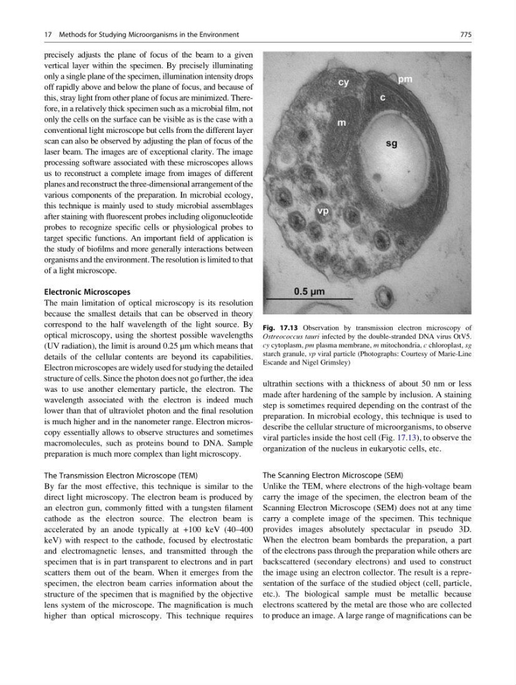
preparation. In microbial ecology, this technique is used to
describe the cellular structure of microorganisms, to observe
viral particles inside the host cell (Fig. 17.13), to observe the
organization of the nucleus in eukaryotic cells, etc.
The Scanning Electron Microscope (SEM)
Unlike the TEM, where electrons of the high-voltage beam
carry the image of the specimen, the electron beam of the
Scanning Electron Microscope (SEM) does not at any time
carry a complete image of the specimen. This technique
provides images absolutely spectacular in pseudo 3D.
When the electron beam bombards the preparation, a part
of the electrons pass through the preparation while others are
backscattered (secondary electrons) and used to construct
the image using an electron collector. The result is a representation of the surface of the studied object (cell, particle,
etc.). The biological sample must be metallic because
electrons scattered by the metal are those who are collected
to produce an image. A large range of magnifications can be
Fig. 17.13 Observation by transmission electron microscopy of
Ostreococcus tauri infected by the double-stranded DNA virus OtV5.
cy cytoplasm, pm plasma membrane, m mitochondria, c chloroplast, sg
starch granule, vp viral particle (Photographs: Courtesy of Marie-Line
Escande and Nigel Grimsley)
17 Methods for Studying Microorganisms in the Environment
775