

fluorescent primary antibody, while the indirect detection
involves binding of a non-fluorescent primary antibody
followed by the addition of a fluorescent secondary antibody
directed against the primary one.
The properties of the antigen-antibody complexes can
also be used to specifically concentrate the targeted
cells by immunocapture. One of the most commonly
immunocapture method used in microbiology is ELISA*
(enzyme-linked immunosorbent assay allowing the quantification of an antibody-antigen reaction from an enzymatic
assay), but its quantitative limits do not allow a reliable
quantification of rare microorganisms. A second immunocapture technique is the immuno-magnetic separation (IMS).
This method uses magnetic beads coated with monoclonal or
polyclonal antibodies. The antibody-antigen reaction allows
efficient separation of the cells from complex matrices. After
this separation, the target microorganisms can be identified
by various techniques such as culture-based or nucleic
acid-based methods.
The major limitations of IF and IMS are linked to the lack
of specificity of the antibodies used in the natural environment and to the absence of physiological information on the
detected cells detected.
15.9.1.3 Enzymatic Methods
The detection of bacterial species in environmental samples
by enzymatic test relies on the detection of a specific enzymatic activity. Such methods were developed and tested
to estimate Escherichia coli counts in wastewaters and
surface waters. These assays are based on the detection of
β-glucuronidase (GUD) activities that can cleave 4methylumbelliferyl β-D-glucuronide (MUG) and release
4-methylumbelliferone (MUF). When exposed to UV light,
MU exhibits a bluish fluorescence that is easily detected.
Over 95 % of the E. coli strains can produce GUD, including
anaerogenic (non-gas-producing) ones. Exceptions are the
enterohemorrhagic E. coli (EHEC) strains of serotype O157:
H7, which are consistently GUD negative.
The measurement of the GUD activity can be achieved
at a global scale (Box 15.6) and a cellular one. As the
fluorescence intensity is directly correlated to the number
of microorganisms in environmental samples, global scale
measurements are used to describe the intensity of the
β-D-glucuronidase activity of a water sample and thus, indirectly, quantify E. coli cells. At the cellular level, these
methods can allow a more accurate detection and quantification of E. coli cells expressing β-D-glucuronidase through an
SPC fluorescence detection.
The estimation of β-D-glucuronidase activities is now
widely used to monitor E. coli among environmental
samples, and several commercial kits making use of the
MUG substrate are now available (Box 15.7).
Box 15.5: DVC-FISH: In Situ Hybridization Coupled with
a Viability Test for the Enumeration of Viable E. coli
Philippe Lebaron
Fluorescent in situ hybridization (FISH, cf. Sect. 15.9.1)
can be coupled with a viability test to specifically detect
specific viable bacteria by microscopy or cytometry. The
direct viable count (DVC) test combines the culture
of the bacteria in a rich medium and the use of antibiotics
that inhibit the formation of the septum and, as a result,
the cell division. After incubation, the viable cells appear
as elongated cells, whereas nonviable cells remain at
their initial size. The viable cell elongation is concomitant with an increase of the RNA content of the cells that
could be easily detected by FISH after staining with a
specific fluorescent molecular probe, without any modification of the total number of bacteria detected. This
combined method has doubled benefits since it allows an
easy enumeration of active cells (showing a metabolic
activity) of a specific species in complex communities
(Garcia Armisen and Servais 2004). This method was
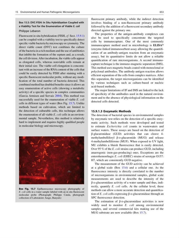
successfully used for the enumeration of viable E. coli
cells in different types of water (Box Fig. 15.7). Unlike
methods based on cultivation, which are limited to
the detection of culturable cells, this technique allows
the enumeration of all viable E. coli cells in an environmental sample. Nevertheless, this method is relatively
hard to implement and requires highly qualified people
in molecular biology and microscopy.
Box Fig. 15.7 Epifluorescence microscopy photography of
E. coli cells in a water sample labeled with an in situ fluorescent
molecular probe (Photograph: Philippe Catala, photograph
collection of Laboratoire Arago, Banyuls)
15 Environmental and Human Pathogenic Microorganisms
653