

15.9.1.1 Fluorescent In Situ Hybridization (FISH)
FISH enables detection of specific nucleic acid sequences
inside intact cells. Probes, oligonucleotides, or peptide nucleic
acids (PNA), complementary to specific sequences in the
cellular DNA or RNA molecules, are combined with
fluorochromes to allow the detection of the hybridized cells
(cf. Sect. 17.7). The main DNA targets are genes (rrs and rrl)
encoding ribosomal RNA (16S rRNA and 23S rRNA).
Although this technique is quite specific and sensitive, it has
limitations when applied to the detection of physiologically
stressed cells or less active ones. This limit can be overcome
by coupling FISH and direct viable count (DVC) (Box 15.5).
15.9.1.2 Immunofluorescence (IF)
Immunofluorescence is based on the formation of a specific
antibody-antigen complex. This method allows the detection, identification, and enumeration of targeted cells in a
complex matrix. Direct detection requires the use of a
Molecular methods
Destructive
Partially quantitative
DIRECT METHODS
INDIRECT METHODS
Culture on specific
media
PCR on specific genes
Real Time PCR
Specific activity detection
using radioactive or
fluorescent probes
(total population)
DNA microarray
Cell detection and quantification
- Epifluorescence microscopy
- Flow cytometry
- Solid-phase cytometry
Cell-based methods
non destructive
quantitative
- in situ hybridization
- Immunofluorescence
- Enzymatic assay (cell-based)
Fluorescent probes
Fig. 15.11 Direct (destructive and nondestructive) and indirect methods used for the detection and quantification of pathogenic microorganisms
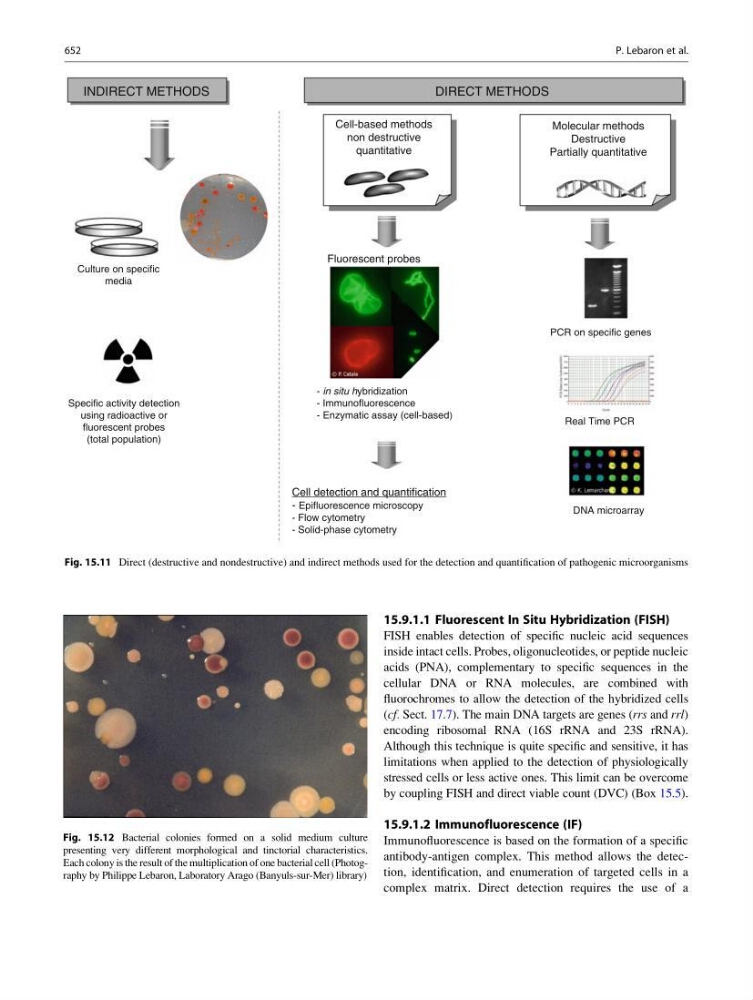
Fig. 15.12 Bacterial colonies formed on a solid medium culture
presenting very different morphological and tinctorial characteristics.
Each colony is the result of the multiplication of one bacterial cell (Photography by Philippe Lebaron, Laboratory Arago (Banyuls-sur-Mer) library)
652
P. Lebaron et al.