

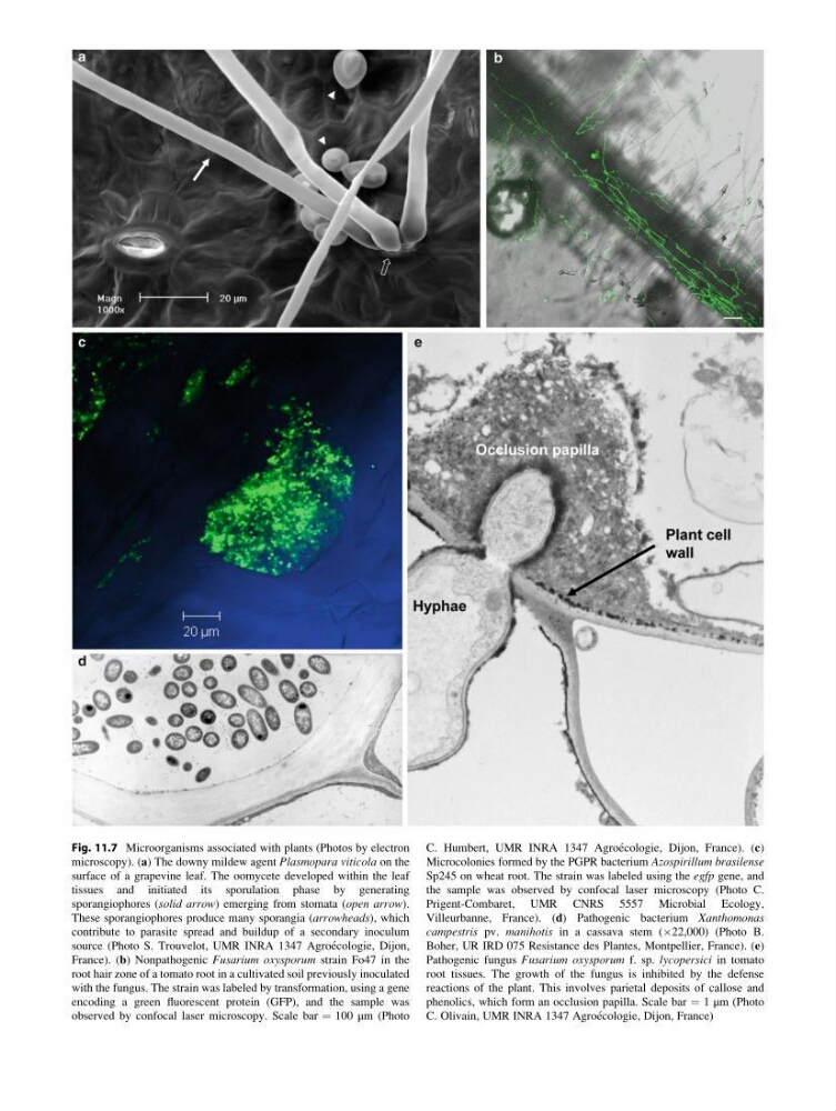
Fig. 11.7 Microorganisms associated with plants (Photos by electron
microscopy). (a) The downy mildew agent Plasmopara viticola on the
surface of a grapevine leaf. The oomycete developed within the leaf
tissues and initiated its sporulation phase by generating
sporangiophores (solid arrow) emerging from stomata (open arrow).
These sporangiophores produce many sporangia (arrowheads), which
contribute to parasite spread and buildup of a secondary inoculum
source (Photo S. Trouvelot, UMR INRA 1347 Agroe ´cologie, Dijon,
France). (b) Nonpathogenic Fusarium oxysporum strain Fo47 in the
root hair zone of a tomato root in a cultivated soil previously inoculated
with the fungus. The strain was labeled by transformation, using a gene
encoding a green fluorescent protein (GFP), and the sample was
observed by confocal laser microscopy. Scale bar ¼ 100 μm (Photo
C. Humbert, UMR INRA 1347 Agroe ´cologie, Dijon, France). (c)
Microcolonies formed by the PGPR bacterium Azospirillum brasilense
Sp245 on wheat root. The strain was labeled using the egfp gene, and
the sample was observed by confocal laser microscopy (Photo C.
Prigent-Combaret, UMR CNRS 5557 Microbial Ecology,
Villeurbanne, France). (d) Pathogenic bacterium Xanthomonas
campestris pv. manihotis in a cassava stem (Â22,000) (Photo B.
Boher, UR IRD 075 Resistance des Plantes, Montpellier, France). (e)
Pathogenic fungus Fusarium oxysporum f. sp. lycopersici in tomato
root tissues. The growth of the fungus is inhibited by the defense
reactions of the plant. This involves parietal deposits of callose and
phenolics, which form an occlusion papilla. Scale bar ¼ 1 μm (Photo
C. Olivain, UMR INRA 1347 Agroe ´cologie, Dijon, France)
microscopy). (a) The downy mildew agent Plasmopara viticola on the
surface of a grapevine leaf. The oomycete developed within the leaf
tissues and initiated its sporulation phase by generating
sporangiophores (solid arrow) emerging from stomata (open arrow).
These sporangiophores produce many sporangia (arrowheads), which
contribute to parasite spread and buildup of a secondary inoculum
source (Photo S. Trouvelot, UMR INRA 1347 Agroe ´cologie, Dijon,
France). (b) Nonpathogenic Fusarium oxysporum strain Fo47 in the
root hair zone of a tomato root in a cultivated soil previously inoculated
with the fungus. The strain was labeled by transformation, using a gene
encoding a green fluorescent protein (GFP), and the sample was
observed by confocal laser microscopy. Scale bar ¼ 100 μm (Photo
C. Humbert, UMR INRA 1347 Agroe ´cologie, Dijon, France). (c)
Microcolonies formed by the PGPR bacterium Azospirillum brasilense
Sp245 on wheat root. The strain was labeled using the egfp gene, and
the sample was observed by confocal laser microscopy (Photo C.
Prigent-Combaret, UMR CNRS 5557 Microbial Ecology,
Villeurbanne, France). (d) Pathogenic bacterium Xanthomonas
campestris pv. manihotis in a cassava stem (Â22,000) (Photo B.
Boher, UR IRD 075 Resistance des Plantes, Montpellier, France). (e)
Pathogenic fungus Fusarium oxysporum f. sp. lycopersici in tomato
root tissues. The growth of the fungus is inhibited by the defense
reactions of the plant. This involves parietal deposits of callose and
phenolics, which form an occlusion papilla. Scale bar ¼ 1 μm (Photo
C. Olivain, UMR INRA 1347 Agroe ´cologie, Dijon, France)