

(CSP “cold-shock proteins”) found in archaea and bacteria.
These proteins have a high affinity for RNA.
The gene domain involved in the attachment to the RNA
is now called the CSP domain; it has been found in many
proteins involved in different functions: adaptation to low
temperatures, cell growth, nutritional stress, and stationary
phase (Graumann and Marahiel 1998).
9.2.4 Protein Glycosylation
Protein glycosylation in eukaryotes is mainly a means
of cellular addressing of proteins to the cell membrane,
for it to be incorporated into it or to be secreted. The
presence of glycosylated motifs is also a way to modulate
the biochemical properties and activities of many proteins
and is dependent on posttranslational modifications.
If many cases of glycosylation exist in archaea, it is
only recently that some cases have been described in
bacteria, especially in relation to virulence (Abu-Qarn
et al. 2008).
Glycosylation enzyme binds specific saccharides to
residues, thus altering their structure and function. These
saccharides are attached to different parts of a few amino
acids, mainly asparagine (N-glycosylation of the amide
function), serine, threonine (O-glycosylation of the
hydroxyl-oxygen), etc.
Several aspects of the physiology of proteins are
influenced by glycosylation, in particular adhesion, recognition, stability, and folding. They are also important
mediators in different processes of signaling and targeting.
In the metazoan pathogen, Campylobacter jejuni, a
heptasaccharide (Table 9.5) is assembled through the
activity of five glycosyltransferases starting with a membrane lipid carrier intermediate (“lipid carrier protein”),
galactose derivatives, and N-acetyl-glucosamine before it
passes through the membrane and is transferred as a whole
to an asparagine residue of a protein outside the membrane.
The mutants for this biosynthetic pathway have an
attenuated phenotype (Abu-Qarn et al. 2008).
In the Proteobacteria Neisseria spp. and Pseudomonas
aeruginosa, pili contain O-glycosylated proteins. In
Neisseria spp., a trisaccharide composed of galactose and
of the unusual 4-diacetamido-2,4,6-trideoxyhexose residue
is attached to a conserved serine of the pilin. In Pseudomonas and Helicobacter, an unusual residue is also present on
the pilin, pseudaminic acid.
Given these saccharides appear to be important for pilus
assembly and virulence; they constitute targets for new
drugs searches.
nitrogen
in excess
nitrogen
deficiency
U
U
U
A
A
A
A
A
GlnB
GS
UTase
UTr
Glu + NH4
Gln
GlnB-U
GS-A
AmtB
AmtR
NAG kinase
PipX
NifA
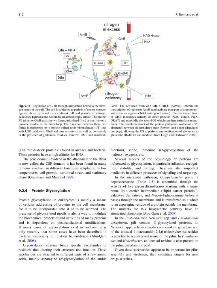
Fig. 9.15 Regulation of GlnB through uridylation linked to the nitrogen status of the cell. The cell is subjected to periods of excess nitrogen
figured above by a red cursor almost full and periods of nitrogen
deficiency figured at the bottom by an almost empty cursor. The protein
PII trimer or GlnB exists in two forms, uridylated (low) or not (top) on a
tyrosine residue of the outer loop. The transition between these two
forms is performed by a protein called uridylyltransferase (UT) that
adds UTP residues to GlnB and thus activates it as well or, conversely
in the presence of glutamine residues, removes UMP and inactivate
GlnB. The activated form of GlnB, GlnB-U (bottom), inhibits the
transcription of repressor AmtR (and activate transport of ammonium)
and activates regulator NifA (nitrogen fixation). The inactivated form
of GlnB modulates activity of other proteins (NAG kinase, PipX,
DRAT) and especially the adenyl GS which can then assimilate ammonium. The double hexamer of the protein glutamine synthetase (GS)
alternates between an adenylated state (bottom) and a non-adenylated
one (top), allowing the GS to perform ammonification of glutamate to
glutamine (Redrawn and modified from Leigh and Dodsworth 2007)
312
P. Normand et al.
These proteins have a high affinity for RNA.
The gene domain involved in the attachment to the RNA
is now called the CSP domain; it has been found in many
proteins involved in different functions: adaptation to low
temperatures, cell growth, nutritional stress, and stationary
phase (Graumann and Marahiel 1998).
9.2.4 Protein Glycosylation
Protein glycosylation in eukaryotes is mainly a means
of cellular addressing of proteins to the cell membrane,
for it to be incorporated into it or to be secreted. The
presence of glycosylated motifs is also a way to modulate
the biochemical properties and activities of many proteins
and is dependent on posttranslational modifications.
If many cases of glycosylation exist in archaea, it is
only recently that some cases have been described in
bacteria, especially in relation to virulence (Abu-Qarn
et al. 2008).
Glycosylation enzyme binds specific saccharides to
residues, thus altering their structure and function. These
saccharides are attached to different parts of a few amino
acids, mainly asparagine (N-glycosylation of the amide
function), serine, threonine (O-glycosylation of the
hydroxyl-oxygen), etc.
Several aspects of the physiology of proteins are
influenced by glycosylation, in particular adhesion, recognition, stability, and folding. They are also important
mediators in different processes of signaling and targeting.
In the metazoan pathogen, Campylobacter jejuni, a
heptasaccharide (Table 9.5) is assembled through the
activity of five glycosyltransferases starting with a membrane lipid carrier intermediate (“lipid carrier protein”),
galactose derivatives, and N-acetyl-glucosamine before it
passes through the membrane and is transferred as a whole
to an asparagine residue of a protein outside the membrane.
The mutants for this biosynthetic pathway have an
attenuated phenotype (Abu-Qarn et al. 2008).
In the Proteobacteria Neisseria spp. and Pseudomonas
aeruginosa, pili contain O-glycosylated proteins. In
Neisseria spp., a trisaccharide composed of galactose and
of the unusual 4-diacetamido-2,4,6-trideoxyhexose residue
is attached to a conserved serine of the pilin. In Pseudomonas and Helicobacter, an unusual residue is also present on
the pilin, pseudaminic acid.
Given these saccharides appear to be important for pilus
assembly and virulence; they constitute targets for new
drugs searches.
nitrogen
in excess
nitrogen
deficiency
U
U
U
A
A
A
A
A
GlnB
GS
UTase
UTr
Glu + NH4
Gln
GlnB-U
GS-A
AmtB
AmtR
NAG kinase
PipX
NifA
Fig. 9.15 Regulation of GlnB through uridylation linked to the nitrogen status of the cell. The cell is subjected to periods of excess nitrogen
figured above by a red cursor almost full and periods of nitrogen
deficiency figured at the bottom by an almost empty cursor. The protein
PII trimer or GlnB exists in two forms, uridylated (low) or not (top) on a
tyrosine residue of the outer loop. The transition between these two
forms is performed by a protein called uridylyltransferase (UT) that
adds UTP residues to GlnB and thus activates it as well or, conversely
in the presence of glutamine residues, removes UMP and inactivate
GlnB. The activated form of GlnB, GlnB-U (bottom), inhibits the
transcription of repressor AmtR (and activate transport of ammonium)
and activates regulator NifA (nitrogen fixation). The inactivated form
of GlnB modulates activity of other proteins (NAG kinase, PipX,
DRAT) and especially the adenyl GS which can then assimilate ammonium. The double hexamer of the protein glutamine synthetase (GS)
alternates between an adenylated state (bottom) and a non-adenylated
one (top), allowing the GS to perform ammonification of glutamate to
glutamine (Redrawn and modified from Leigh and Dodsworth 2007)
312
P. Normand et al.