

from this organism and used in reverse genetics to identify
the corresponding genes. Analysis of these genes has
highlighted two conserved domains, called GGDEF and
EAL (named based on the conserved amino acid residues),
that were found in other bacterial proteins. Phylogenetic
analysis of genes encoding GGDEF domains or EAL
shows they are widely distributed in bacteria but absent
from archaea and eukaryotes, suggesting that the di-cGMP
is limited to the bacterial world. The biochemical study of
many GGDEF domain proteins has shown that this domain
catalyzed the synthesis of di-cGMP from two GTPs
(Fig. 9.12) (Ryjenkov et al. 2005).
The di-cGMP is hydrolyzed to 5
0 -pGpG by the EAL
domain of PDEA. Recently, another protein domain, HDGYP, whose phylogenetic distribution is similar to that of
GGDEF and EAL domains, capable of directly hydrolyzing
di-cGMP into two GMP, was described. The reasons for the
presence of two systems of hydrolysis of di-cGMP in the
same genome are still unknown. Most proteins containing
GGDEF and EAL domains have a modular structure
combining to these two areas, other conserved domains
involved in both perception (MASE, CACHE, PAS, GAF)
and in transduction (REC) and signal response (HTH).
The GGDEF and EAL domains are often found in tandem
in the same protein, but usually only one enzyme activity is
expressed in vivo by the native protein (Fig. 9.13).
The structural diversity of proteins with GGDEF and EAL
domains contributes to a fine-level regulation of the concentration of di-cGMP in the cell. Bacteria with a large number
of GGDEF and EAL domain proteins have in common the
ability to survive in a wide variety of environmental media.
For example, Pseudomonas aeruginosa encodes 17 GGDEF
domain proteins, 5 with an EAL domain, 16 with a GGDEFEAL domain, and 3 with an HD-GYP domain; Vibrio
cholerae in turn encodes 31 GGDEF domain proteins, 22
with an EAL domain, 10 with a GGDEF-EAL domain, and 9
with an HD-GYP-domain.
Various works on the physiological role of di-cGMP have
made it clear that the di-cGMP activates biofilm formation
and inhibits cell motility, thus regulating the transition
between planktonic and sessile states of bacterial cells.
The fimX mutation of P. aeruginosa, a gene that encodes
a PDEA (Fig. 9.13), leads to an increase in the intracellular
concentration of di-cGMP and an absence of type IV pili on
the cell surface with a consequent loss of twitching motility
(Kazmierczak et al. 2006). Also in P. aeruginosa, constitutive activation of WspR (Fig. 9.13), the response regulator
with a DGC activity of the MCP receptor chemosensor
Wsp, causes motility inhibition and activation of biofilm
formation (Hickman et al. 2005). The decrease in flagellar
motility has been demonstrated in Salmonella enterica
serovar typhimurium and Vibrio cholerae. In Salmonella,
ectopic expression of AdrA, a DGC (Fig. 9.13) leads to an
inhibition of motility, the opposite effect being observed
with YhjH, a PDEA (Simm et al. 2004). Similar experiments
CheA
CheA
CheW
CheW
CheZ
MCP
MCP
MCP
MCP
Flagellar motor basis
Outer membrane
Cytoplasmic
membrane
Peptidoglycan (wall)
Periplasm
Cytoplasm
CheB
CheB
P
P
P
O-CH 3
CH 3
CH 3
CheY
CheY
CheR
+
-
PBP
Flagellum
Cytoplasmic
membrane
-C=
O
C
O
HO
E
=
_
_
E
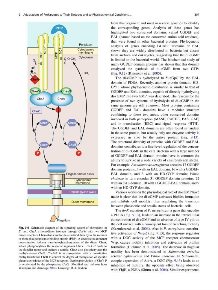
Fig. 9.9 Schematic diagram of the signaling system of chemotaxis in
E. coli. CheA a homodimer interacts through CheW with two MCP
dimer receptors. Chemotactic molecules can bind directly to the receiver
or through a periplasmic binding protein (PBP). A decrease in attractant
concentration induces trans-autophosphorylation of the dimer CheA,
which phosphorylates the response regulator CheY. CheY-P binds to
the flagellar motor and induces a tumble. CheA also phosphorylates the
methylesterase CheB. CheB-P is in competition with a constitutive
methyltransferase CheR to control the degree of methylation of specific
glutamate residues of the MCP receptors. Dephosphorylation of CheY-P
is accelerated by the phosphatase CheZ (Modified and redrawn from
Wadhams and Armitage 2004). Drawing: M.-J. Bodiou
9 Adaptations of Prokaryotes to Their Biotopes and to Physicochemical Conditions. . .
307
the corresponding genes. Analysis of these genes has
highlighted two conserved domains, called GGDEF and
EAL (named based on the conserved amino acid residues),
that were found in other bacterial proteins. Phylogenetic
analysis of genes encoding GGDEF domains or EAL
shows they are widely distributed in bacteria but absent
from archaea and eukaryotes, suggesting that the di-cGMP
is limited to the bacterial world. The biochemical study of
many GGDEF domain proteins has shown that this domain
catalyzed the synthesis of di-cGMP from two GTPs
(Fig. 9.12) (Ryjenkov et al. 2005).
The di-cGMP is hydrolyzed to 5
0 -pGpG by the EAL
domain of PDEA. Recently, another protein domain, HDGYP, whose phylogenetic distribution is similar to that of
GGDEF and EAL domains, capable of directly hydrolyzing
di-cGMP into two GMP, was described. The reasons for the
presence of two systems of hydrolysis of di-cGMP in the
same genome are still unknown. Most proteins containing
GGDEF and EAL domains have a modular structure
combining to these two areas, other conserved domains
involved in both perception (MASE, CACHE, PAS, GAF)
and in transduction (REC) and signal response (HTH).
The GGDEF and EAL domains are often found in tandem
in the same protein, but usually only one enzyme activity is
expressed in vivo by the native protein (Fig. 9.13).
The structural diversity of proteins with GGDEF and EAL
domains contributes to a fine-level regulation of the concentration of di-cGMP in the cell. Bacteria with a large number
of GGDEF and EAL domain proteins have in common the
ability to survive in a wide variety of environmental media.
For example, Pseudomonas aeruginosa encodes 17 GGDEF
domain proteins, 5 with an EAL domain, 16 with a GGDEFEAL domain, and 3 with an HD-GYP domain; Vibrio
cholerae in turn encodes 31 GGDEF domain proteins, 22
with an EAL domain, 10 with a GGDEF-EAL domain, and 9
with an HD-GYP-domain.
Various works on the physiological role of di-cGMP have
made it clear that the di-cGMP activates biofilm formation
and inhibits cell motility, thus regulating the transition
between planktonic and sessile states of bacterial cells.
The fimX mutation of P. aeruginosa, a gene that encodes
a PDEA (Fig. 9.13), leads to an increase in the intracellular
concentration of di-cGMP and an absence of type IV pili on
the cell surface with a consequent loss of twitching motility
(Kazmierczak et al. 2006). Also in P. aeruginosa, constitutive activation of WspR (Fig. 9.13), the response regulator
with a DGC activity of the MCP receptor chemosensor
Wsp, causes motility inhibition and activation of biofilm
formation (Hickman et al. 2005). The decrease in flagellar
motility has been demonstrated in Salmonella enterica
serovar typhimurium and Vibrio cholerae. In Salmonella,
ectopic expression of AdrA, a DGC (Fig. 9.13) leads to an
inhibition of motility, the opposite effect being observed
with YhjH, a PDEA (Simm et al. 2004). Similar experiments
CheA
CheA
CheW
CheW
CheZ
MCP
MCP
MCP
MCP
Flagellar motor basis
Outer membrane
Cytoplasmic
membrane
Peptidoglycan (wall)
Periplasm
Cytoplasm
CheB
CheB
P
P
P
O-CH 3
CH 3
CH 3
CheY
CheY
CheR
+
-
PBP
Flagellum
Cytoplasmic
membrane
-C=
O
C
O
HO
E
=
_
_
E
Fig. 9.9 Schematic diagram of the signaling system of chemotaxis in
E. coli. CheA a homodimer interacts through CheW with two MCP
dimer receptors. Chemotactic molecules can bind directly to the receiver
or through a periplasmic binding protein (PBP). A decrease in attractant
concentration induces trans-autophosphorylation of the dimer CheA,
which phosphorylates the response regulator CheY. CheY-P binds to
the flagellar motor and induces a tumble. CheA also phosphorylates the
methylesterase CheB. CheB-P is in competition with a constitutive
methyltransferase CheR to control the degree of methylation of specific
glutamate residues of the MCP receptors. Dephosphorylation of CheY-P
is accelerated by the phosphatase CheZ (Modified and redrawn from
Wadhams and Armitage 2004). Drawing: M.-J. Bodiou
9 Adaptations of Prokaryotes to Their Biotopes and to Physicochemical Conditions. . .
307