

other monomer. CheY and CheB compete for binding to P2.
CheA transfers phosphoryl groups faster to CheY than
to CheB, which allows triggering of the response before
adaptation of the MCP receiver.
Chemotactic molecules bind to the periplasmic domain of
MCPs at the interface between two monomers. Many MCPs
are capable of binding two different ligands, such as the Tar
receptor of E. coli, which binds directly aspartate and,
through a periplasmic binding protein (PBP), maltose.
MCPs are predominantly localized to one pole of the cell,
where they form clusters involving several thousands of
receptors (Fig. 9.11). E. coli has five MCPs, Tar, Tsr (serine), Trg (ribose, galactose), Tap (dipeptides), and Aer, a
particular MCP involved in aerotactism. Aer lacks a periplasmic domain but has at its end N-terminal a cytoplasmic
PAS domain that complexes FAD whose oxidation state is
related to changes in oxygen concentration of the medium
(Fig. 9.4).
Tar and Tsr are present at about 3,000 copies per cell,
while Trg, Tap, and Aer at a few hundred. Although the
precise molecular architecture of these clusters is not
established, it is clear that interactions between cytoplasmic
domains of receptors are crucial for the formation of clusters
and their operation. This cluster organization involving
different MCP receptors creates a network capable of
amplifying allosteric stimuli of different chemoeffectors
before transmission of the signal to other proteins of the
signaling system (Kentner and Sourjik 2006).
9.2.2.4 Ser/Thr/Tyr Kinases (STYKs)
Protein kinase systems play a key role in the signaling
mechanisms in eukaryotic cells. The tyrosine protein kinase
receptors are an important class of membrane receptors,
and serine/threonine protein kinases are involved in the
regulation of many metabolic pathways. Systematic analysis
of sequenced bacterial genomes has permitted to reveal the
presence of numerous proteins with Ser/Thr/Tyr kinase
domains in all bacterial phyla (Krupa and Srinivasan 2005).
The fact that many of these proteins have transmembrane
helices suggests that many of them are membrane receptors
capable of binding extracellular ligands. Thus, STK PknB of
Mycobacterium bovis, for which counterparts are present in
different groups of Gram-positive bacteria, has an extracellular PASTA domain able to fix the D-alanyl-D-alanine
dipeptide of the peptidoglycan and the β-lactam ring of
penicillins. Stimulatory molecules as well as the cellular
targets of bacterial STYKs are poorly known. The membrane STK, AfsK of Streptomyces coelicolor phosphorylates
a global regulator of the secondary metabolism, AfsR.
Another membrane STK of Mycobacterium tuberculosis,
PknH phosphorylates EmbR, a transcription regulator of
the gene-coding arabinosyltransferase, an enzyme involved
in the synthesis of arabinogalactan, a key molecule of the
cell wall of mycobacteria. Various studies have shown the
involvement of STYKs in the formation of fruiting bodies in
Myxococcus xanthus and the development of aerial hyphae
in Streptomyces coelicolor.
9.2.2.5 Second Messenger Systems
Systems with di-cGMP
Di-cGMP is a secondary messenger found in most bacteria.
It regulates many cellular processes including motility,
biofilm formation, and expression of virulence genes
(Tamayo et al. 2007). Di-cGMP was first identified as an
allosteric activator of cellulose synthase in Gluconacetobacter xylinus. The diguanylate cyclase (DGC) and phosphodiesterase A (PDEA), enzymes catalyzing the synthesis
and the degradation of di-cGMP, respectively, were purified
Repulsive
conditions
Attractive
conditions
Tumble
Swiming
Swiming
a
b
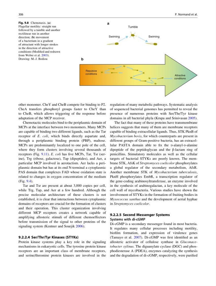
Fig. 9.8 Chemotaxis. (a)
Flagellar motility: straight run
followed by a tumble and another
rectilinear run in another
direction; (b) movement
of a bacterium in a gradient
of attractant with longer strokes
in the direction of attractive
conditions (Modified and redrawn
from Webre et al. 2003).
Drawing: M.-J. Bodiou
306
P. Normand et al.
CheA transfers phosphoryl groups faster to CheY than
to CheB, which allows triggering of the response before
adaptation of the MCP receiver.
Chemotactic molecules bind to the periplasmic domain of
MCPs at the interface between two monomers. Many MCPs
are capable of binding two different ligands, such as the Tar
receptor of E. coli, which binds directly aspartate and,
through a periplasmic binding protein (PBP), maltose.
MCPs are predominantly localized to one pole of the cell,
where they form clusters involving several thousands of
receptors (Fig. 9.11). E. coli has five MCPs, Tar, Tsr (serine), Trg (ribose, galactose), Tap (dipeptides), and Aer, a
particular MCP involved in aerotactism. Aer lacks a periplasmic domain but has at its end N-terminal a cytoplasmic
PAS domain that complexes FAD whose oxidation state is
related to changes in oxygen concentration of the medium
(Fig. 9.4).
Tar and Tsr are present at about 3,000 copies per cell,
while Trg, Tap, and Aer at a few hundred. Although the
precise molecular architecture of these clusters is not
established, it is clear that interactions between cytoplasmic
domains of receptors are crucial for the formation of clusters
and their operation. This cluster organization involving
different MCP receptors creates a network capable of
amplifying allosteric stimuli of different chemoeffectors
before transmission of the signal to other proteins of the
signaling system (Kentner and Sourjik 2006).
9.2.2.4 Ser/Thr/Tyr Kinases (STYKs)
Protein kinase systems play a key role in the signaling
mechanisms in eukaryotic cells. The tyrosine protein kinase
receptors are an important class of membrane receptors,
and serine/threonine protein kinases are involved in the
regulation of many metabolic pathways. Systematic analysis
of sequenced bacterial genomes has permitted to reveal the
presence of numerous proteins with Ser/Thr/Tyr kinase
domains in all bacterial phyla (Krupa and Srinivasan 2005).
The fact that many of these proteins have transmembrane
helices suggests that many of them are membrane receptors
capable of binding extracellular ligands. Thus, STK PknB of
Mycobacterium bovis, for which counterparts are present in
different groups of Gram-positive bacteria, has an extracellular PASTA domain able to fix the D-alanyl-D-alanine
dipeptide of the peptidoglycan and the β-lactam ring of
penicillins. Stimulatory molecules as well as the cellular
targets of bacterial STYKs are poorly known. The membrane STK, AfsK of Streptomyces coelicolor phosphorylates
a global regulator of the secondary metabolism, AfsR.
Another membrane STK of Mycobacterium tuberculosis,
PknH phosphorylates EmbR, a transcription regulator of
the gene-coding arabinosyltransferase, an enzyme involved
in the synthesis of arabinogalactan, a key molecule of the
cell wall of mycobacteria. Various studies have shown the
involvement of STYKs in the formation of fruiting bodies in
Myxococcus xanthus and the development of aerial hyphae
in Streptomyces coelicolor.
9.2.2.5 Second Messenger Systems
Systems with di-cGMP
Di-cGMP is a secondary messenger found in most bacteria.
It regulates many cellular processes including motility,
biofilm formation, and expression of virulence genes
(Tamayo et al. 2007). Di-cGMP was first identified as an
allosteric activator of cellulose synthase in Gluconacetobacter xylinus. The diguanylate cyclase (DGC) and phosphodiesterase A (PDEA), enzymes catalyzing the synthesis
and the degradation of di-cGMP, respectively, were purified
Repulsive
conditions
Attractive
conditions
Tumble
Swiming
Swiming
a
b
Fig. 9.8 Chemotaxis. (a)
Flagellar motility: straight run
followed by a tumble and another
rectilinear run in another
direction; (b) movement
of a bacterium in a gradient
of attractant with longer strokes
in the direction of attractive
conditions (Modified and redrawn
from Webre et al. 2003).
Drawing: M.-J. Bodiou
306
P. Normand et al.