

bacterium perceives conditions that may be harmful, conversely the number of flips decreases, and the bacterium
stays its course when chemical conditions improve, allowing
the bacterium to climb down gradients of repellents concentration and climb up gradients of attractants.
The molecular mechanism of chemotaxis is complex and
depends on the existence of transmembrane receptors called
MCPs (“methyl-accepting chemotaxis proteins”), a specific
histidine protein kinase (CheA), two response regulators
(CheY and CheB, an RR with a methylesterase activity),
and a constitutive methyltransferase (CheR), which allows
methylation from S-adenosylmethionine of glutamate
residues of the MCP receptor (Wadhams and Armitage
2004).
Binding of an attractive or repulsive molecule to the
receptor will change the autophosphorylation activity of
the normal histidine protein kinase CheA, coupled to the
receiver via an adapter protein CheW. The formation of
CheA-P allows the phosphorylation of the response regulator CheY. It is the phosphorylated form of the response
regulator CheY-P that induces a change in the direction of
rotation of the flagellum by binding to the flagellar motor.
In E. coli (Fig. 9.9), a decrease in attractant concentration
in the medium reduces the number of attractant molecules
bound on the MCP receiver, which in turn stimulates the
activity of autophosphorylation of CheA and therefore the
amount of intracellular CheY-P with, as a consequence,
an increase in the frequency of tumbles. One phosphatase,
CheZ, increases the speed of spontaneous dephosphorylation
of CheY-P and ensures a quick stop of the signal.
CheA-P also phosphorylates CheB, but at a lower speed,
resulting in increased methylesterase activity and thus
demethylation of the MCP receiver. When demethylated,
the MCP receiver has a lower capacity to induce phosphorylation of CheA for the same concentration of attractant.
Thus, the rate of autophosphorylation of CheA and the
frequency of tumbles return to the level preceding the stimulation. The system is then adapted and able to receive any
further increase or decrease in the number of bound ligands.
Conversely, an increase in the concentration of attractant
inhibits the autophosphorylation of CheA, which will
decrease the concentration of CheY-P and, ultimately,
allow runs to occur for longer periods in a straight line.
Phosphorylation of CheB and thus its methylesterase activity
will also be reduced, which allows the constitutive CheR
methyltransferase to increase the rate of methylation of
MCPs. When highly methylated, for a given concentration
of attractant, MCP receptors will have a higher capacity to
stimulate autophosphorylation of CheA, bringing the system
to the level of activity preceding the stimulation despite
the continued presence of attractant, leading to a normal
frequency of tumbles.
MCPs are transmembrane homodimers with a periplasmic effector-binding domain, between two transmembrane
helices, a HAMP domain for signal transduction at a highly
conserved cytoplasmic signaling domain (AD) (Fig. 9.10).
MA has sites for methylation/demethylation that are glutamate residues that allow cells to adapt the response of
receptors to a change in concentration of the chemoattractant
over time, as well as a binding site for the adapter protein
CheW and the histidine kinase CheA. This site is also
involved in the association of clusters of MCP receptors.
CheA is a homodimer that has a structure different from
HKs encountered in conventional two-component systems
(Fig. 9.10). Each monomer is indeed made up of five
domains: P1 is a Hpt domain carrying the active His, P2 a
binding domain for CheY and CheB, P3 a dimerization
domain, P4 is a HATPase catalytic domain, and P5 is a
domain coupling CheA to CheW and to the MCP receiver.
Phosphorylation of the His residue of the P1 domain of one
of the monomers is done by the P4 catalytic domain of the
l ll
l
ll
HATPase
HATPase
HisKA / DHp
H
H
ATP
ATP
Histidine kinase
HAMP
Membrane
Sensor
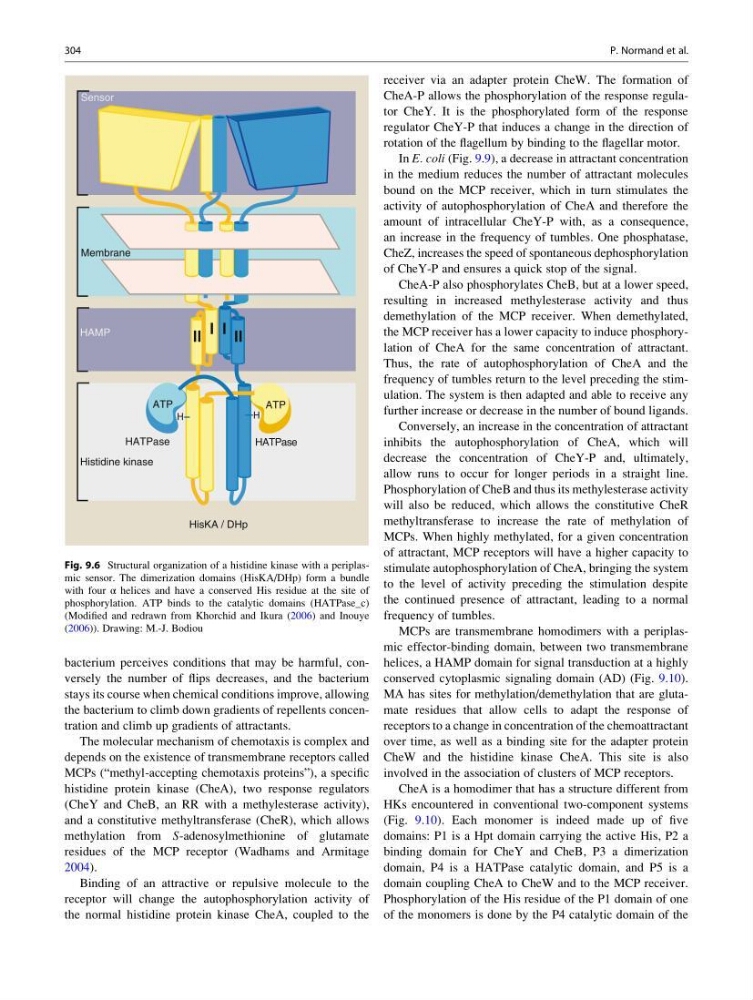
Fig. 9.6 Structural organization of a histidine kinase with a periplasmic sensor. The dimerization domains (HisKA/DHp) form a bundle
with four α helices and have a conserved His residue at the site of
phosphorylation. ATP binds to the catalytic domains (HATPase_c)
(Modified and redrawn from Khorchid and Ikura (2006) and Inouye
(2006)). Drawing: M.-J. Bodiou
304
P. Normand et al.
stays its course when chemical conditions improve, allowing
the bacterium to climb down gradients of repellents concentration and climb up gradients of attractants.
The molecular mechanism of chemotaxis is complex and
depends on the existence of transmembrane receptors called
MCPs (“methyl-accepting chemotaxis proteins”), a specific
histidine protein kinase (CheA), two response regulators
(CheY and CheB, an RR with a methylesterase activity),
and a constitutive methyltransferase (CheR), which allows
methylation from S-adenosylmethionine of glutamate
residues of the MCP receptor (Wadhams and Armitage
2004).
Binding of an attractive or repulsive molecule to the
receptor will change the autophosphorylation activity of
the normal histidine protein kinase CheA, coupled to the
receiver via an adapter protein CheW. The formation of
CheA-P allows the phosphorylation of the response regulator CheY. It is the phosphorylated form of the response
regulator CheY-P that induces a change in the direction of
rotation of the flagellum by binding to the flagellar motor.
In E. coli (Fig. 9.9), a decrease in attractant concentration
in the medium reduces the number of attractant molecules
bound on the MCP receiver, which in turn stimulates the
activity of autophosphorylation of CheA and therefore the
amount of intracellular CheY-P with, as a consequence,
an increase in the frequency of tumbles. One phosphatase,
CheZ, increases the speed of spontaneous dephosphorylation
of CheY-P and ensures a quick stop of the signal.
CheA-P also phosphorylates CheB, but at a lower speed,
resulting in increased methylesterase activity and thus
demethylation of the MCP receiver. When demethylated,
the MCP receiver has a lower capacity to induce phosphorylation of CheA for the same concentration of attractant.
Thus, the rate of autophosphorylation of CheA and the
frequency of tumbles return to the level preceding the stimulation. The system is then adapted and able to receive any
further increase or decrease in the number of bound ligands.
Conversely, an increase in the concentration of attractant
inhibits the autophosphorylation of CheA, which will
decrease the concentration of CheY-P and, ultimately,
allow runs to occur for longer periods in a straight line.
Phosphorylation of CheB and thus its methylesterase activity
will also be reduced, which allows the constitutive CheR
methyltransferase to increase the rate of methylation of
MCPs. When highly methylated, for a given concentration
of attractant, MCP receptors will have a higher capacity to
stimulate autophosphorylation of CheA, bringing the system
to the level of activity preceding the stimulation despite
the continued presence of attractant, leading to a normal
frequency of tumbles.
MCPs are transmembrane homodimers with a periplasmic effector-binding domain, between two transmembrane
helices, a HAMP domain for signal transduction at a highly
conserved cytoplasmic signaling domain (AD) (Fig. 9.10).
MA has sites for methylation/demethylation that are glutamate residues that allow cells to adapt the response of
receptors to a change in concentration of the chemoattractant
over time, as well as a binding site for the adapter protein
CheW and the histidine kinase CheA. This site is also
involved in the association of clusters of MCP receptors.
CheA is a homodimer that has a structure different from
HKs encountered in conventional two-component systems
(Fig. 9.10). Each monomer is indeed made up of five
domains: P1 is a Hpt domain carrying the active His, P2 a
binding domain for CheY and CheB, P3 a dimerization
domain, P4 is a HATPase catalytic domain, and P5 is a
domain coupling CheA to CheW and to the MCP receiver.
Phosphorylation of the His residue of the P1 domain of one
of the monomers is done by the P4 catalytic domain of the
l ll
l
ll
HATPase
HATPase
HisKA / DHp
H
H
ATP
ATP
Histidine kinase
HAMP
Membrane
Sensor
Fig. 9.6 Structural organization of a histidine kinase with a periplasmic sensor. The dimerization domains (HisKA/DHp) form a bundle
with four α helices and have a conserved His residue at the site of
phosphorylation. ATP binds to the catalytic domains (HATPase_c)
(Modified and redrawn from Khorchid and Ikura (2006) and Inouye
(2006)). Drawing: M.-J. Bodiou
304
P. Normand et al.