

(e.g.,
Aureococcus,
Aureoumbra,
Pelagomonas),
Chrysophyceae (e.g., Picophagus), and Pinguiophyceae
(e.g., Pingiococcus) (Fuller et al. 2006).
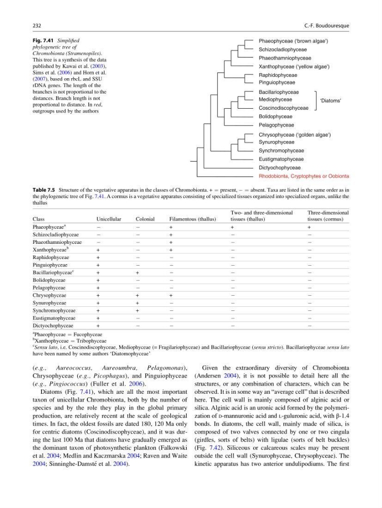
Diatoms (Fig. 7.41), which are all the most important
taxon of unicellular Chromobionta, both by the number of
species and by the role they play in the global primary
production, are relatively recent at the scale of geological
times. In fact, the oldest fossils are dated 180, 120 Ma only
for centric diatoms (Coscinodiscophyceae), and it was during the last 100 Ma that diatoms have gradually emerged as
the dominant taxon of photosynthetic plankton (Falkowski
et al. 2004; Medlin and Kaczmarska 2004; Raven and Waite
2004; Sinninghe-Damste ´ et al. 2004).
Given the extraordinary diversity of Chromobionta
(Andersen 2004), it is not possible to detail here all the
structures, or any combination of characters, which can be
observed. It is in some way an “average cell” that is described
here. The cell wall is mainly composed of alginic acid or
silica. Alginic acid is an uronic acid formed by the polymerization of D-mannuronic acid and L-guluronic acid, with β-1.4
bonds. In diatoms, the cell wall, mainly made of silica, is
composed of two valves connected by one or two cingula
(girdles, sorts of belts) with ligulae (sorts of belt buckles)
(Fig. 7.42). Siliceous or calcareous scales may be present
outside the cell wall (Synurophyceae, Chrysophyceae). The
kinetic apparatus has two anterior undulipodiums. The first
Table 7.5 Structure of the vegetative apparatus in the classes of Chromobionta. + ¼ present, À ¼ absent. Taxa are listed in the same order as in
the phylogenetic tree of Fig. 7.41. A cormus is a vegetative apparatus consisting of specialized tissues organized into specialized organs, unlike the
thallus
Class
Unicellular
Colonial
Filamentous (thallus)
Two- and three-dimensional
tissues (thallus)
Three-dimensional
tissues (cormus)
Phaeophyceae
a
À
À
+
+
+
Schizocladiophyceae
À
À
+
À
À
Phaeothamniophyceae
À
À
+
À
À
Xanthophyceae
b
+
À
+
À
À
Raphidophyceae
+
À
À
À
À
Pinguiophyceae
+
À
À
À
À
Bacillariophyceae
c
+
+
À
À
À
Bolidophyceae
+
À
À
À
À
Pelagophyceae
+
À
À
À
À
Chrysophyceae
+
+
+
À
À
Synurophyceae
+
+
À
À
À
Synchromophyceae
+
+
À
À
À
Eustigmatophyceae
+
À
À
À
À
Dictyochophyceae
+
À
À
À
À
a
Phaeophyceae ¼ Fucophyceae
b
Xanthophyceae ¼ Tribophyceae
c
Sensu lato, i.e. Coscinodiscophyceae, Mediophyceae (= Fragilariophyceae) and Bacillariophyceae (sensu stricto). Bacillariophyceae sensu lato
have been named by some authors ‘Diatomophyceae’
Dictyochophyceae
Eustigmatophyceae
Synchromophyceae
Bolidophyceae
Pelagophyceae
Chrysophyceae (‘golden algae’)
Synurophyceae
Coscinodiscophyceae
Mediophyceae
Bacillariophyceae
Pinguiophyceae
Raphidophyceae
Xanthophyceae (‘yellow algae’)
Rhodobionta, Cryptophytes or Oobionta
Phaeothamniophyceae
Schizocladiophyceae
Phaeophyceae (‘brown algae’)
‘Diatoms’
Fig. 7.41 Simplified
phylogenetic tree of
Chromobionta (Stramenopiles).
This tree is a synthesis of the data
published by Kawai et al. (2003),
Sims et al. (2006) and Horn et al.
(2007), based on rbcL and SSU
rDNA genes. The length of the
branches is not proportional to the
distances. Branch length is not
proportional to distance. In red,
outgroups used by the authors
232
C.-F. Boudouresque
Aureococcus,
Aureoumbra,
Pelagomonas),
Chrysophyceae (e.g., Picophagus), and Pinguiophyceae
(e.g., Pingiococcus) (Fuller et al. 2006).
Diatoms (Fig. 7.41), which are all the most important
taxon of unicellular Chromobionta, both by the number of
species and by the role they play in the global primary
production, are relatively recent at the scale of geological
times. In fact, the oldest fossils are dated 180, 120 Ma only
for centric diatoms (Coscinodiscophyceae), and it was during the last 100 Ma that diatoms have gradually emerged as
the dominant taxon of photosynthetic plankton (Falkowski
et al. 2004; Medlin and Kaczmarska 2004; Raven and Waite
2004; Sinninghe-Damste ´ et al. 2004).
Given the extraordinary diversity of Chromobionta
(Andersen 2004), it is not possible to detail here all the
structures, or any combination of characters, which can be
observed. It is in some way an “average cell” that is described
here. The cell wall is mainly composed of alginic acid or
silica. Alginic acid is an uronic acid formed by the polymerization of D-mannuronic acid and L-guluronic acid, with β-1.4
bonds. In diatoms, the cell wall, mainly made of silica, is
composed of two valves connected by one or two cingula
(girdles, sorts of belts) with ligulae (sorts of belt buckles)
(Fig. 7.42). Siliceous or calcareous scales may be present
outside the cell wall (Synurophyceae, Chrysophyceae). The
kinetic apparatus has two anterior undulipodiums. The first
Table 7.5 Structure of the vegetative apparatus in the classes of Chromobionta. + ¼ present, À ¼ absent. Taxa are listed in the same order as in
the phylogenetic tree of Fig. 7.41. A cormus is a vegetative apparatus consisting of specialized tissues organized into specialized organs, unlike the
thallus
Class
Unicellular
Colonial
Filamentous (thallus)
Two- and three-dimensional
tissues (thallus)
Three-dimensional
tissues (cormus)
Phaeophyceae
a
À
À
+
+
+
Schizocladiophyceae
À
À
+
À
À
Phaeothamniophyceae
À
À
+
À
À
Xanthophyceae
b
+
À
+
À
À
Raphidophyceae
+
À
À
À
À
Pinguiophyceae
+
À
À
À
À
Bacillariophyceae
c
+
+
À
À
À
Bolidophyceae
+
À
À
À
À
Pelagophyceae
+
À
À
À
À
Chrysophyceae
+
+
+
À
À
Synurophyceae
+
+
À
À
À
Synchromophyceae
+
+
À
À
À
Eustigmatophyceae
+
À
À
À
À
Dictyochophyceae
+
À
À
À
À
a
Phaeophyceae ¼ Fucophyceae
b
Xanthophyceae ¼ Tribophyceae
c
Sensu lato, i.e. Coscinodiscophyceae, Mediophyceae (= Fragilariophyceae) and Bacillariophyceae (sensu stricto). Bacillariophyceae sensu lato
have been named by some authors ‘Diatomophyceae’
Dictyochophyceae
Eustigmatophyceae
Synchromophyceae
Bolidophyceae
Pelagophyceae
Chrysophyceae (‘golden algae’)
Synurophyceae
Coscinodiscophyceae
Mediophyceae
Bacillariophyceae
Pinguiophyceae
Raphidophyceae
Xanthophyceae (‘yellow algae’)
Rhodobionta, Cryptophytes or Oobionta
Phaeothamniophyceae
Schizocladiophyceae
Phaeophyceae (‘brown algae’)
‘Diatoms’
Fig. 7.41 Simplified
phylogenetic tree of
Chromobionta (Stramenopiles).
This tree is a synthesis of the data
published by Kawai et al. (2003),
Sims et al. (2006) and Horn et al.
(2007), based on rbcL and SSU
rDNA genes. The length of the
branches is not proportional to the
distances. Branch length is not
proportional to distance. In red,
outgroups used by the authors
232
C.-F. Boudouresque