

off California at a depth of ~600 m (in the aphotic zone); it
retains chloroplasts derived from diatoms closely related to
Skeletonema costatum and Odontella sinensis, species which
live in the photic pelagic zone; the chloroplasts are probably
from fecal pellets of zooplanktonic organisms which fall to
the bottom; they could be used for the assimilation of inorganic nitrogen, meeting the nitrogen requirements of the
host (Grzymski et al. 2002).
7.7
Super-Kingdom Chromalveolata
The kingdoms Alveolata and Stramenopiles have a number
of common characteristics. It seems that the monophyly of
this super-group is strong. They are therefore referred to as
Chromalveolata. Some insertae sedis taxa, such as
Haptobionta, could belong to this super-group (Fig. 7.1).
Some authors, e.g. Palmer et al. (2004), consider that a
single endosymbiosis founding event was at the origin of the
chloroplast in Chromalveolata. This secondary endosymbiosis with a Rhodobionta would have occurred in the common
ancestor of all Chromalveolata (cf. Sect. 5.4). According to
this hypothesis, the absence of photosynthesis in taxa such as
Oobionta, Labyrinthulobionta, and ciliates should be due to
a secondary loss. The existence of a single founding event in
the photosynthesis of Chromalveolata is however disputed
(e.g., Bodyl et al. 2009).
The Chromalveolata have a number of shared
characters. They mainly concern the photosynthetic taxa.
(i) Mitochondria have cristae
15 of the tubular type. (ii)
Chlorophyll a is associated with chlorophyll c (c1, c2,
and/or c3). (iii) The thylakoids are never isolated, but
stacked into two- or three-layered lamellae. (iv) The
chloroplasts (where present) are surrounded by a fourmembrane envelope: the classical two-membrane envelope
and an outer second two-membrane envelope. The latter
constitutes the remains of the secondary endosymbiosis.
This outer envelope (the periplastidial envelope) may (or
not) be confluent with the nuclear envelope. It should be
noted that these characters, taken in isolation, are not specific to Chromalveolata.
7.8
Kingdom Alveolata
7.8.1 General Remarks
The kingdom Alveolata (¼alveolate, Alveolobionta) is a
taxon strongly supported by molecular phylogenies. The
most notable shared characteristic is the presence of flattened
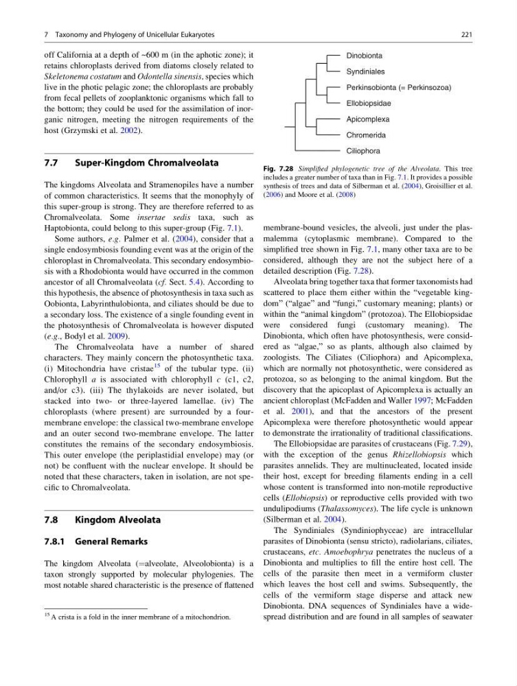
membrane-bound vesicles, the alveoli, just under the plasmalemma (cytoplasmic membrane). Compared to the
simplified tree shown in Fig. 7.1, many other taxa are to be
considered, although they are not the subject here of a
detailed description (Fig. 7.28).
Alveolata bring together taxa that former taxonomists had
scattered to place them either within the “vegetable kingdom” (“algae” and “fungi,” customary meaning; plants) or
within the “animal kingdom” (protozoa). The Ellobiopsidae
were considered fungi (customary meaning). The
Dinobionta, which often have photosynthesis, were considered as “algae,” so as plants, although also claimed by
zoologists. The Ciliates (Ciliophora) and Apicomplexa,
which are normally not photosynthetic, were considered as
protozoa, so as belonging to the animal kingdom. But the
discovery that the apicoplast of Apicomplexa is actually an
ancient chloroplast (McFadden and Waller 1997; McFadden
et al. 2001), and that the ancestors of the present
Apicomplexa were therefore photosynthetic would appear
to demonstrate the irrationality of traditional classifications.
The Ellobiopsidae are parasites of crustaceans (Fig. 7.29),
with the exception of the genus Rhizellobiopsis which
parasites annelids. They are multinucleated, located inside
their host, except for breeding filaments ending in a cell
whose content is transformed into non-motile reproductive
cells (Ellobiopsis) or reproductive cells provided with two
undulipodiums (Thalassomyces). The life cycle is unknown
(Silberman et al. 2004).
The Syndiniales (Syndiniophyceae) are intracellular
parasites of Dinobionta (sensu stricto), radiolarians, ciliates,
crustaceans, etc. Amoebophrya penetrates the nucleus of a
Dinobionta and multiplies to fill the entire host cell. The
cells of the parasite then meet in a vermiform cluster
which leaves the host cell and swims. Subsequently, the
cells of the vermiform stage disperse and attack new
Dinobionta. DNA sequences of Syndiniales have a widespread distribution and are found in all samples of seawater
Ciliophora
Chromerida
Apicomplexa
Ellobiopsidae
Perkinsobionta (= Perkinsozoa)
Syndiniales
Dinobionta
Fig. 7.28 Simplified phylogenetic tree of the Alveolata. This tree
includes a greater number of taxa than in Fig. 7.1. It provides a possible
synthesis of trees and data of Silberman et al. (2004), Groisillier et al.
(2006) and Moore et al. (2008)
15 A crista is a fold in the inner membrane of a mitochondrion.
7 Taxonomy and Phylogeny of Unicellular Eukaryotes
221
retains chloroplasts derived from diatoms closely related to
Skeletonema costatum and Odontella sinensis, species which
live in the photic pelagic zone; the chloroplasts are probably
from fecal pellets of zooplanktonic organisms which fall to
the bottom; they could be used for the assimilation of inorganic nitrogen, meeting the nitrogen requirements of the
host (Grzymski et al. 2002).
7.7
Super-Kingdom Chromalveolata
The kingdoms Alveolata and Stramenopiles have a number
of common characteristics. It seems that the monophyly of
this super-group is strong. They are therefore referred to as
Chromalveolata. Some insertae sedis taxa, such as
Haptobionta, could belong to this super-group (Fig. 7.1).
Some authors, e.g. Palmer et al. (2004), consider that a
single endosymbiosis founding event was at the origin of the
chloroplast in Chromalveolata. This secondary endosymbiosis with a Rhodobionta would have occurred in the common
ancestor of all Chromalveolata (cf. Sect. 5.4). According to
this hypothesis, the absence of photosynthesis in taxa such as
Oobionta, Labyrinthulobionta, and ciliates should be due to
a secondary loss. The existence of a single founding event in
the photosynthesis of Chromalveolata is however disputed
(e.g., Bodyl et al. 2009).
The Chromalveolata have a number of shared
characters. They mainly concern the photosynthetic taxa.
(i) Mitochondria have cristae
15 of the tubular type. (ii)
Chlorophyll a is associated with chlorophyll c (c1, c2,
and/or c3). (iii) The thylakoids are never isolated, but
stacked into two- or three-layered lamellae. (iv) The
chloroplasts (where present) are surrounded by a fourmembrane envelope: the classical two-membrane envelope
and an outer second two-membrane envelope. The latter
constitutes the remains of the secondary endosymbiosis.
This outer envelope (the periplastidial envelope) may (or
not) be confluent with the nuclear envelope. It should be
noted that these characters, taken in isolation, are not specific to Chromalveolata.
7.8
Kingdom Alveolata
7.8.1 General Remarks
The kingdom Alveolata (¼alveolate, Alveolobionta) is a
taxon strongly supported by molecular phylogenies. The
most notable shared characteristic is the presence of flattened
membrane-bound vesicles, the alveoli, just under the plasmalemma (cytoplasmic membrane). Compared to the
simplified tree shown in Fig. 7.1, many other taxa are to be
considered, although they are not the subject here of a
detailed description (Fig. 7.28).
Alveolata bring together taxa that former taxonomists had
scattered to place them either within the “vegetable kingdom” (“algae” and “fungi,” customary meaning; plants) or
within the “animal kingdom” (protozoa). The Ellobiopsidae
were considered fungi (customary meaning). The
Dinobionta, which often have photosynthesis, were considered as “algae,” so as plants, although also claimed by
zoologists. The Ciliates (Ciliophora) and Apicomplexa,
which are normally not photosynthetic, were considered as
protozoa, so as belonging to the animal kingdom. But the
discovery that the apicoplast of Apicomplexa is actually an
ancient chloroplast (McFadden and Waller 1997; McFadden
et al. 2001), and that the ancestors of the present
Apicomplexa were therefore photosynthetic would appear
to demonstrate the irrationality of traditional classifications.
The Ellobiopsidae are parasites of crustaceans (Fig. 7.29),
with the exception of the genus Rhizellobiopsis which
parasites annelids. They are multinucleated, located inside
their host, except for breeding filaments ending in a cell
whose content is transformed into non-motile reproductive
cells (Ellobiopsis) or reproductive cells provided with two
undulipodiums (Thalassomyces). The life cycle is unknown
(Silberman et al. 2004).
The Syndiniales (Syndiniophyceae) are intracellular
parasites of Dinobionta (sensu stricto), radiolarians, ciliates,
crustaceans, etc. Amoebophrya penetrates the nucleus of a
Dinobionta and multiplies to fill the entire host cell. The
cells of the parasite then meet in a vermiform cluster
which leaves the host cell and swims. Subsequently, the
cells of the vermiform stage disperse and attack new
Dinobionta. DNA sequences of Syndiniales have a widespread distribution and are found in all samples of seawater
Ciliophora
Chromerida
Apicomplexa
Ellobiopsidae
Perkinsobionta (= Perkinsozoa)
Syndiniales
Dinobionta
Fig. 7.28 Simplified phylogenetic tree of the Alveolata. This tree
includes a greater number of taxa than in Fig. 7.1. It provides a possible
synthesis of trees and data of Silberman et al. (2004), Groisillier et al.
(2006) and Moore et al. (2008)
15 A crista is a fold in the inner membrane of a mitochondrion.
7 Taxonomy and Phylogeny of Unicellular Eukaryotes
221