

Box 7.1 (continued)
diverged early in the history of Eukaryota: the
picobiliphytes (Not et al. 2007) and the rappemonades
(Kim et al. 2011b). Usually, environmental lineages
could be placed within known taxonomic groups such
as Fungi, Archaeplastida, Rhizaria, Stramenopiles, or
even associated to finer taxonomic levels. The discovery
of widely distributed environmental lineages not only
helps refine the Eukaryotic tree of Life but also provides
a measure of the gap in our knowledge of eukaryotic
biodiversity. Metagenetic surveys of micro-Eukaryotes
revealed that many novel taxa are far from being marginal or rare and therefore may represent, a substantial
fraction of the total microbial biomass. For example,
diverse stramenopile phyla referred to as MAST (MArine
STramenopiles) were identified all across the world’s
oceans, except for the MAST-4 group in polar areas
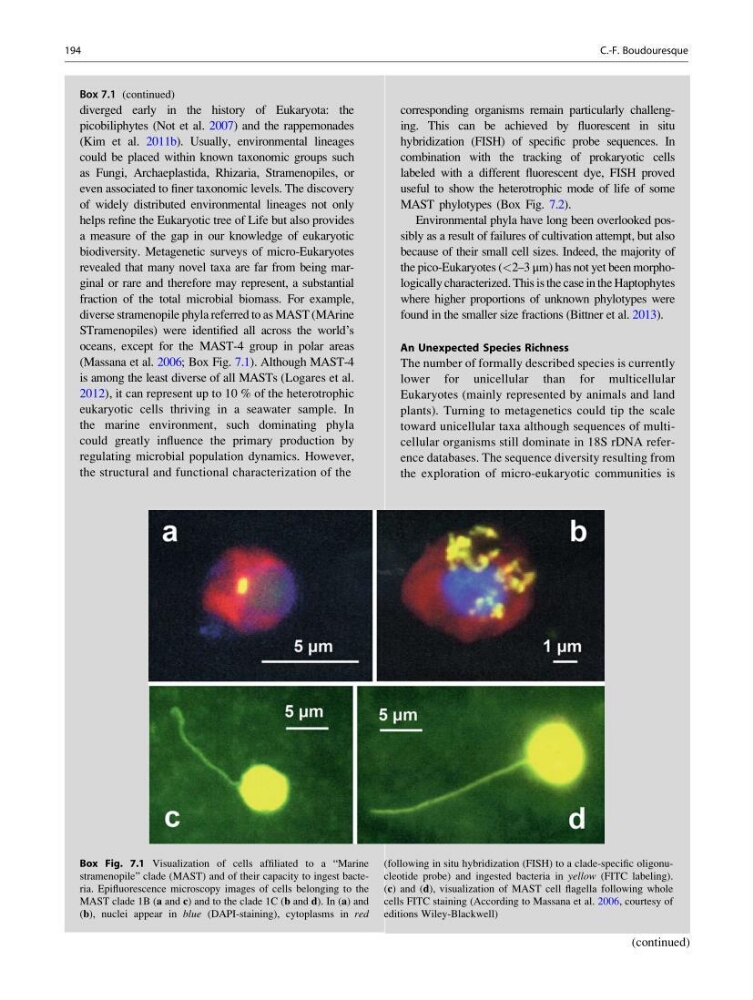
(Massana et al. 2006; Box Fig. 7.1). Although MAST-4
is among the least diverse of all MASTs (Logares et al.
2012), it can represent up to 10 % of the heterotrophic
eukaryotic cells thriving in a seawater sample. In
the marine environment, such dominating phyla
could greatly influence the primary production by
regulating microbial population dynamics. However,
the structural and functional characterization of the
(continued)
Box 7.1 (continued)
corresponding organisms remain particularly challenging. This can be achieved by fluorescent in situ
hybridization (FISH) of specific probe sequences. In
combination with the tracking of prokaryotic cells
labeled with a different fluorescent dye, FISH proved
useful to show the heterotrophic mode of life of some
MAST phylotypes (Box Fig. 7.2).
Environmental phyla have long been overlooked possibly as a result of failures of cultivation attempt, but also
because of their small cell sizes. Indeed, the majority of
the pico-Eukaryotes (<2–3 μm) has not yet been morphologically characterized. This is the case in the Haptophytes
where higher proportions of unknown phylotypes were
found in the smaller size fractions (Bittner et al. 2013).
An Unexpected Species Richness
The number of formally described species is currently
lower for unicellular than for multicellular
Eukaryotes (mainly represented by animals and land
plants). Turning to metagenetics could tip the scale
toward unicellular taxa although sequences of multicellular organisms still dominate in 18S rDNA reference databases. The sequence diversity resulting from
the exploration of micro-eukaryotic communities is
(continued)
Box Fig. 7.1 Visualization of cells affiliated to a “Marine
stramenopile” clade (MAST) and of their capacity to ingest bacteria. Epifluorescence microscopy images of cells belonging to the
MAST clade 1B (a and c) and to the clade 1C (b and d). In (a) and
(b), nuclei appear in blue (DAPI-staining), cytoplasms in red
(following in situ hybridization (FISH) to a clade-specific oligonucleotide probe) and ingested bacteria in yellow (FITC labeling).
(c) and (d), visualization of MAST cell flagella following whole
cells FITC staining (According to Massana et al. 2006, courtesy of
editions Wiley-Blackwell)
194
C.-F. Boudouresque
diverged early in the history of Eukaryota: the
picobiliphytes (Not et al. 2007) and the rappemonades
(Kim et al. 2011b). Usually, environmental lineages
could be placed within known taxonomic groups such
as Fungi, Archaeplastida, Rhizaria, Stramenopiles, or
even associated to finer taxonomic levels. The discovery
of widely distributed environmental lineages not only
helps refine the Eukaryotic tree of Life but also provides
a measure of the gap in our knowledge of eukaryotic
biodiversity. Metagenetic surveys of micro-Eukaryotes
revealed that many novel taxa are far from being marginal or rare and therefore may represent, a substantial
fraction of the total microbial biomass. For example,
diverse stramenopile phyla referred to as MAST (MArine
STramenopiles) were identified all across the world’s
oceans, except for the MAST-4 group in polar areas
(Massana et al. 2006; Box Fig. 7.1). Although MAST-4
is among the least diverse of all MASTs (Logares et al.
2012), it can represent up to 10 % of the heterotrophic
eukaryotic cells thriving in a seawater sample. In
the marine environment, such dominating phyla
could greatly influence the primary production by
regulating microbial population dynamics. However,
the structural and functional characterization of the
(continued)
Box 7.1 (continued)
corresponding organisms remain particularly challenging. This can be achieved by fluorescent in situ
hybridization (FISH) of specific probe sequences. In
combination with the tracking of prokaryotic cells
labeled with a different fluorescent dye, FISH proved
useful to show the heterotrophic mode of life of some
MAST phylotypes (Box Fig. 7.2).
Environmental phyla have long been overlooked possibly as a result of failures of cultivation attempt, but also
because of their small cell sizes. Indeed, the majority of
the pico-Eukaryotes (<2–3 μm) has not yet been morphologically characterized. This is the case in the Haptophytes
where higher proportions of unknown phylotypes were
found in the smaller size fractions (Bittner et al. 2013).
An Unexpected Species Richness
The number of formally described species is currently
lower for unicellular than for multicellular
Eukaryotes (mainly represented by animals and land
plants). Turning to metagenetics could tip the scale
toward unicellular taxa although sequences of multicellular organisms still dominate in 18S rDNA reference databases. The sequence diversity resulting from
the exploration of micro-eukaryotic communities is
(continued)
Box Fig. 7.1 Visualization of cells affiliated to a “Marine
stramenopile” clade (MAST) and of their capacity to ingest bacteria. Epifluorescence microscopy images of cells belonging to the
MAST clade 1B (a and c) and to the clade 1C (b and d). In (a) and
(b), nuclei appear in blue (DAPI-staining), cytoplasms in red
(following in situ hybridization (FISH) to a clade-specific oligonucleotide probe) and ingested bacteria in yellow (FITC labeling).
(c) and (d), visualization of MAST cell flagella following whole
cells FITC staining (According to Massana et al. 2006, courtesy of
editions Wiley-Blackwell)
194
C.-F. Boudouresque