

(T ¼ A) and three hydrogen bonds between the G and C
bases (G C). The calculation of G + C% is
G þ C% ¼
G þ C mol  100
Mol G þ C
ð
Þþ A þ T
ð
Þ
ð
Þ
:
Due to the wide range of G + C percentages in genomic
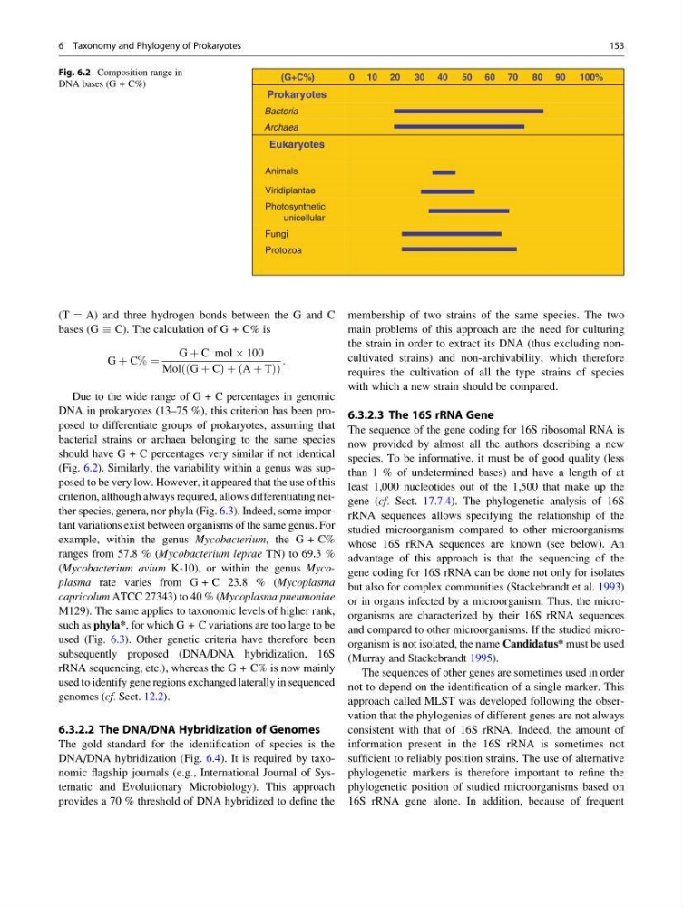
DNA in prokaryotes (13–75 %), this criterion has been proposed to differentiate groups of prokaryotes, assuming that
bacterial strains or archaea belonging to the same species
should have G + C percentages very similar if not identical
(Fig. 6.2). Similarly, the variability within a genus was supposed to be very low. However, it appeared that the use of this
criterion, although always required, allows differentiating neither species, genera, nor phyla (Fig. 6.3). Indeed, some important variations exist between organisms of the same genus. For
example, within the genus Mycobacterium, the G + C%
ranges from 57.8 % (Mycobacterium leprae TN) to 69.3 %
(Mycobacterium avium K-10), or within the genus Mycoplasma rate varies from G + C 23.8 % (Mycoplasma
capricolum ATCC 27343) to 40 % (Mycoplasma pneumoniae
M129). The same applies to taxonomic levels of higher rank,
such as phyla*, for which G + C variations are too large to be
used (Fig. 6.3). Other genetic criteria have therefore been
subsequently proposed (DNA/DNA hybridization, 16S
rRNA sequencing, etc.), whereas the G + C% is now mainly
used to identify gene regions exchanged laterally in sequenced
genomes (cf. Sect. 12.2).
6.3.2.2 The DNA/DNA Hybridization of Genomes
The gold standard for the identification of species is the
DNA/DNA hybridization (Fig. 6.4). It is required by taxonomic flagship journals (e.g., International Journal of Systematic and Evolutionary Microbiology). This approach
provides a 70 % threshold of DNA hybridized to define the
membership of two strains of the same species. The two
main problems of this approach are the need for culturing
the strain in order to extract its DNA (thus excluding noncultivated strains) and non-archivability, which therefore
requires the cultivation of all the type strains of species
with which a new strain should be compared.
6.3.2.3 The 16S rRNA Gene
The sequence of the gene coding for 16S ribosomal RNA is
now provided by almost all the authors describing a new
species. To be informative, it must be of good quality (less
than 1 % of undetermined bases) and have a length of at
least 1,000 nucleotides out of the 1,500 that make up the
gene (cf. Sect. 17.7.4). The phylogenetic analysis of 16S
rRNA sequences allows specifying the relationship of the
studied microorganism compared to other microorganisms
whose 16S rRNA sequences are known (see below). An
advantage of this approach is that the sequencing of the
gene coding for 16S rRNA can be done not only for isolates
but also for complex communities (Stackebrandt et al. 1993)
or in organs infected by a microorganism. Thus, the microorganisms are characterized by their 16S rRNA sequences
and compared to other microorganisms. If the studied microorganism is not isolated, the name Candidatus* must be used
(Murray and Stackebrandt 1995).
The sequences of other genes are sometimes used in order
not to depend on the identification of a single marker. This
approach called MLST was developed following the observation that the phylogenies of different genes are not always
consistent with that of 16S rRNA. Indeed, the amount of
information present in the 16S rRNA is sometimes not
sufficient to reliably position strains. The use of alternative
phylogenetic markers is therefore important to refine the
phylogenetic position of studied microorganisms based on
16S rRNA gene alone. In addition, because of frequent
(G+C%)
0 10 20
30 40
50 60 70
80 90
100%
Prokaryotes
Bacteria
Archaea
Eukaryotes
Animals
Viridiplantae
Photosynthetic
unicellular
Fungi
Protozoa
Fig. 6.2 Composition range in
DNA bases (G + C%)
6 Taxonomy and Phylogeny of Prokaryotes
153
bases (G C). The calculation of G + C% is
G þ C% ¼
G þ C mol  100
Mol G þ C
ð
Þþ A þ T
ð
Þ
ð
Þ
:
Due to the wide range of G + C percentages in genomic
DNA in prokaryotes (13–75 %), this criterion has been proposed to differentiate groups of prokaryotes, assuming that
bacterial strains or archaea belonging to the same species
should have G + C percentages very similar if not identical
(Fig. 6.2). Similarly, the variability within a genus was supposed to be very low. However, it appeared that the use of this
criterion, although always required, allows differentiating neither species, genera, nor phyla (Fig. 6.3). Indeed, some important variations exist between organisms of the same genus. For
example, within the genus Mycobacterium, the G + C%
ranges from 57.8 % (Mycobacterium leprae TN) to 69.3 %
(Mycobacterium avium K-10), or within the genus Mycoplasma rate varies from G + C 23.8 % (Mycoplasma
capricolum ATCC 27343) to 40 % (Mycoplasma pneumoniae
M129). The same applies to taxonomic levels of higher rank,
such as phyla*, for which G + C variations are too large to be
used (Fig. 6.3). Other genetic criteria have therefore been
subsequently proposed (DNA/DNA hybridization, 16S
rRNA sequencing, etc.), whereas the G + C% is now mainly
used to identify gene regions exchanged laterally in sequenced
genomes (cf. Sect. 12.2).
6.3.2.2 The DNA/DNA Hybridization of Genomes
The gold standard for the identification of species is the
DNA/DNA hybridization (Fig. 6.4). It is required by taxonomic flagship journals (e.g., International Journal of Systematic and Evolutionary Microbiology). This approach
provides a 70 % threshold of DNA hybridized to define the
membership of two strains of the same species. The two
main problems of this approach are the need for culturing
the strain in order to extract its DNA (thus excluding noncultivated strains) and non-archivability, which therefore
requires the cultivation of all the type strains of species
with which a new strain should be compared.
6.3.2.3 The 16S rRNA Gene
The sequence of the gene coding for 16S ribosomal RNA is
now provided by almost all the authors describing a new
species. To be informative, it must be of good quality (less
than 1 % of undetermined bases) and have a length of at
least 1,000 nucleotides out of the 1,500 that make up the
gene (cf. Sect. 17.7.4). The phylogenetic analysis of 16S
rRNA sequences allows specifying the relationship of the
studied microorganism compared to other microorganisms
whose 16S rRNA sequences are known (see below). An
advantage of this approach is that the sequencing of the
gene coding for 16S rRNA can be done not only for isolates
but also for complex communities (Stackebrandt et al. 1993)
or in organs infected by a microorganism. Thus, the microorganisms are characterized by their 16S rRNA sequences
and compared to other microorganisms. If the studied microorganism is not isolated, the name Candidatus* must be used
(Murray and Stackebrandt 1995).
The sequences of other genes are sometimes used in order
not to depend on the identification of a single marker. This
approach called MLST was developed following the observation that the phylogenies of different genes are not always
consistent with that of 16S rRNA. Indeed, the amount of
information present in the 16S rRNA is sometimes not
sufficient to reliably position strains. The use of alternative
phylogenetic markers is therefore important to refine the
phylogenetic position of studied microorganisms based on
16S rRNA gene alone. In addition, because of frequent
(G+C%)
0 10 20
30 40
50 60 70
80 90
100%
Prokaryotes
Bacteria
Archaea
Eukaryotes
Animals
Viridiplantae
Photosynthetic
unicellular
Fungi
Protozoa
Fig. 6.2 Composition range in
DNA bases (G + C%)
6 Taxonomy and Phylogeny of Prokaryotes
153