

“Asian Sea,” was resolved by designation of a neotype
from central Indonesia (Wallace, 1999). The size of the
genus was reduced slightly by elevation of a subgenus
Isopora to separate genus status because it differed from
other Acropora in skeletal and reproductive morphology
as well as reproductive mode (Wallace et al., 2007). The
remaining species are organized into 20 species groups
based on skeletal features (Veron and Wallace, 1984;
Wallace, 1999). Genetic studies are revealing numerous
dilemmas about species boundaries (Van Oppen et al.,
2001, 2002) and at least one named species of Acropora
is now known to be an F1 hybrid (Van Oppen et al.,
2000; Vollmer and Palumbi, 2002).
The skeleton and polyps
All scleractinian corals have skeletons of the crystal aragonite form of calcium carbonate, but formation of
a skeleton by the polyps follows different patterns among
families, giving structural features by which corals can be
identified in both living and fossil form (Wells, 1956;
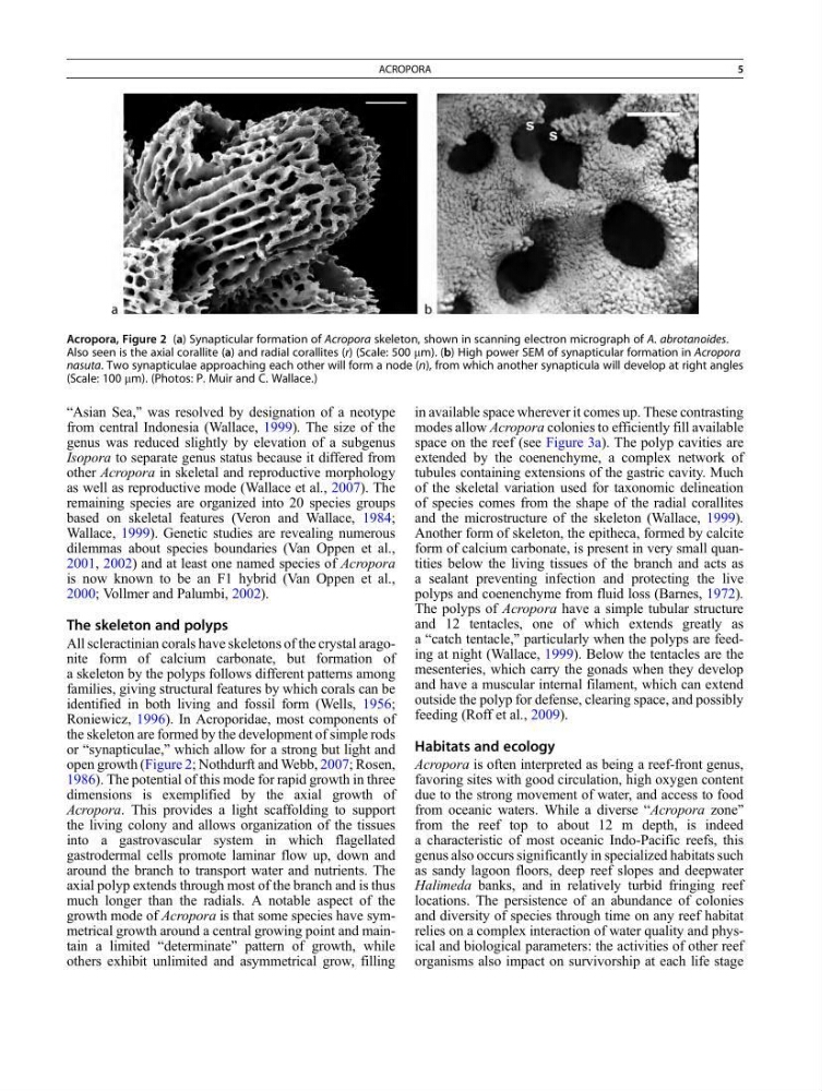
Roniewicz, 1996). In Acroporidae, most components of
the skeleton are formed by the development of simple rods
or “synapticulae,” which allow for a strong but light and
open growth (Figure 2; Nothdurft and Webb, 2007; Rosen,
1986). The potential of this mode for rapid growth in three
dimensions is exemplified by the axial growth of
Acropora. This provides a light scaffolding to support
the living colony and allows organization of the tissues
into a gastrovascular system in which flagellated
gastrodermal cells promote laminar flow up, down and
around the branch to transport water and nutrients. The
axial polyp extends through most of the branch and is thus
much longer than the radials. A notable aspect of the
growth mode of Acropora is that some species have symmetrical growth around a central growing point and maintain a limited “determinate” pattern of growth, while
others exhibit unlimited and asymmetrical grow, filling
in available space wherever it comes up. These contrasting
modes allow Acropora colonies to efficiently fill available
space on the reef (see Figure 3a). The polyp cavities are
extended by the coenenchyme, a complex network of
tubules containing extensions of the gastric cavity. Much
of the skeletal variation used for taxonomic delineation
of species comes from the shape of the radial corallites
and the microstructure of the skeleton (Wallace, 1999).
Another form of skeleton, the epitheca, formed by calcite
form of calcium carbonate, is present in very small quantities below the living tissues of the branch and acts as
a sealant preventing infection and protecting the live
polyps and coenenchyme from fluid loss (Barnes, 1972).
The polyps of Acropora have a simple tubular structure
and 12 tentacles, one of which extends greatly as
a “catch tentacle,” particularly when the polyps are feeding at night (Wallace, 1999). Below the tentacles are the
mesenteries, which carry the gonads when they develop
and have a muscular internal filament, which can extend
outside the polyp for defense, clearing space, and possibly
feeding (Roff et al., 2009).
Habitats and ecology
Acropora is often interpreted as being a reef-front genus,
favoring sites with good circulation, high oxygen content
due to the strong movement of water, and access to food
from oceanic waters. While a diverse “Acropora zone”
from the reef top to about 12 m depth, is indeed
a characteristic of most oceanic Indo-Pacific reefs, this
genus also occurs significantly in specialized habitats such
as sandy lagoon floors, deep reef slopes and deepwater
Halimeda banks, and in relatively turbid fringing reef
locations. The persistence of an abundance of colonies
and diversity of species through time on any reef habitat
relies on a complex interaction of water quality and physical and biological parameters: the activities of other reef
organisms also impact on survivorship at each life stage
Acropora, Figure 2 (a) Synapticular formation of Acropora skeleton, shown in scanning electron micrograph of A. abrotanoides.
Also seen is the axial corallite (a) and radial corallites (r) (Scale: 500 mm). (b) High power SEM of synapticular formation in Acropora
nasuta. Two synapticulae approaching each other will form a node (n), from which another synapticula will develop at right angles
(Scale: 100 mm). (Photos: P. Muir and C. Wallace.)
ACROPORA
5
from central Indonesia (Wallace, 1999). The size of the
genus was reduced slightly by elevation of a subgenus
Isopora to separate genus status because it differed from
other Acropora in skeletal and reproductive morphology
as well as reproductive mode (Wallace et al., 2007). The
remaining species are organized into 20 species groups
based on skeletal features (Veron and Wallace, 1984;
Wallace, 1999). Genetic studies are revealing numerous
dilemmas about species boundaries (Van Oppen et al.,
2001, 2002) and at least one named species of Acropora
is now known to be an F1 hybrid (Van Oppen et al.,
2000; Vollmer and Palumbi, 2002).
The skeleton and polyps
All scleractinian corals have skeletons of the crystal aragonite form of calcium carbonate, but formation of
a skeleton by the polyps follows different patterns among
families, giving structural features by which corals can be
identified in both living and fossil form (Wells, 1956;
Roniewicz, 1996). In Acroporidae, most components of
the skeleton are formed by the development of simple rods
or “synapticulae,” which allow for a strong but light and
open growth (Figure 2; Nothdurft and Webb, 2007; Rosen,
1986). The potential of this mode for rapid growth in three
dimensions is exemplified by the axial growth of
Acropora. This provides a light scaffolding to support
the living colony and allows organization of the tissues
into a gastrovascular system in which flagellated
gastrodermal cells promote laminar flow up, down and
around the branch to transport water and nutrients. The
axial polyp extends through most of the branch and is thus
much longer than the radials. A notable aspect of the
growth mode of Acropora is that some species have symmetrical growth around a central growing point and maintain a limited “determinate” pattern of growth, while
others exhibit unlimited and asymmetrical grow, filling
in available space wherever it comes up. These contrasting
modes allow Acropora colonies to efficiently fill available
space on the reef (see Figure 3a). The polyp cavities are
extended by the coenenchyme, a complex network of
tubules containing extensions of the gastric cavity. Much
of the skeletal variation used for taxonomic delineation
of species comes from the shape of the radial corallites
and the microstructure of the skeleton (Wallace, 1999).
Another form of skeleton, the epitheca, formed by calcite
form of calcium carbonate, is present in very small quantities below the living tissues of the branch and acts as
a sealant preventing infection and protecting the live
polyps and coenenchyme from fluid loss (Barnes, 1972).
The polyps of Acropora have a simple tubular structure
and 12 tentacles, one of which extends greatly as
a “catch tentacle,” particularly when the polyps are feeding at night (Wallace, 1999). Below the tentacles are the
mesenteries, which carry the gonads when they develop
and have a muscular internal filament, which can extend
outside the polyp for defense, clearing space, and possibly
feeding (Roff et al., 2009).
Habitats and ecology
Acropora is often interpreted as being a reef-front genus,
favoring sites with good circulation, high oxygen content
due to the strong movement of water, and access to food
from oceanic waters. While a diverse “Acropora zone”
from the reef top to about 12 m depth, is indeed
a characteristic of most oceanic Indo-Pacific reefs, this
genus also occurs significantly in specialized habitats such
as sandy lagoon floors, deep reef slopes and deepwater
Halimeda banks, and in relatively turbid fringing reef
locations. The persistence of an abundance of colonies
and diversity of species through time on any reef habitat
relies on a complex interaction of water quality and physical and biological parameters: the activities of other reef
organisms also impact on survivorship at each life stage
Acropora, Figure 2 (a) Synapticular formation of Acropora skeleton, shown in scanning electron micrograph of A. abrotanoides.
Also seen is the axial corallite (a) and radial corallites (r) (Scale: 500 mm). (b) High power SEM of synapticular formation in Acropora
nasuta. Two synapticulae approaching each other will form a node (n), from which another synapticula will develop at right angles
(Scale: 100 mm). (Photos: P. Muir and C. Wallace.)
ACROPORA
5