

recently found to be harder (Ma et al. 2008) than plates and fibers. It is clear from
Fig. 7.8a that it is the polycrystalline matrix in the stone part that becomes the sharp,
hard tip of the tooth that does the grinding.
Recently, Ma et al. (2009) reported that the polycrystalline matrix is highly
co-oriented in P. lividus. Furthermore, Robach et al. (2009) demonstrated that
polycrystalline matrix and fibers share the same orientation in L. variegatus,
whereas Killian et al. (2009) and Yang et al. (2011) made the same observation
in S. purpuratus. These concurring observations raise an important question: how
does the polycrystalline matrix form and co-orient its crystal nanoparticles? Are the
nanoparticles aggregating before or after crystallizing? In other words, is this the
result of oriented attachment of crystalline nanoparticles, as first observed by Penn
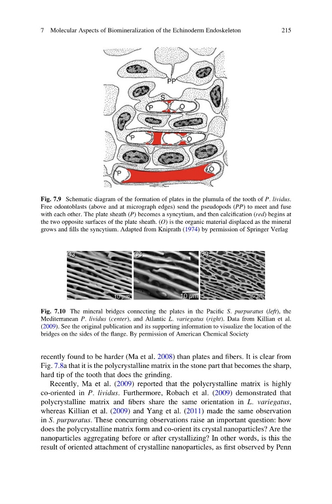
Fig. 7.10 The mineral bridges connecting the plates in the Pacific S. purpuratus (left), the
Mediterranean P. lividus (center), and Atlantic L. variegatus (right). Data from Killian et al.
(2009). See the original publication and its supporting information to visualize the location of the
bridges on the sides of the flange. By permission of American Chemical Society
Fig. 7.9 Schematic diagram of the formation of plates in the plumula of the tooth of P. lividus.
Free odontoblasts (above and at micrograph edges) send the pseudopods (PP) to meet and fuse
with each other. The plate sheath (P) becomes a syncytium, and then calcification (red) begins at
the two opposite surfaces of the plate sheath. (O) is the organic material displaced as the mineral
grows and fills the syncytium. Adapted from Kniprath (1974) by permission of Springer Verlag
7 Molecular Aspects of Biomineralization of the Echinoderm Endoskeleton
215
Fig. 7.8a that it is the polycrystalline matrix in the stone part that becomes the sharp,
hard tip of the tooth that does the grinding.
Recently, Ma et al. (2009) reported that the polycrystalline matrix is highly
co-oriented in P. lividus. Furthermore, Robach et al. (2009) demonstrated that
polycrystalline matrix and fibers share the same orientation in L. variegatus,
whereas Killian et al. (2009) and Yang et al. (2011) made the same observation
in S. purpuratus. These concurring observations raise an important question: how
does the polycrystalline matrix form and co-orient its crystal nanoparticles? Are the
nanoparticles aggregating before or after crystallizing? In other words, is this the
result of oriented attachment of crystalline nanoparticles, as first observed by Penn
Fig. 7.10 The mineral bridges connecting the plates in the Pacific S. purpuratus (left), the
Mediterranean P. lividus (center), and Atlantic L. variegatus (right). Data from Killian et al.
(2009). See the original publication and its supporting information to visualize the location of the
bridges on the sides of the flange. By permission of American Chemical Society
Fig. 7.9 Schematic diagram of the formation of plates in the plumula of the tooth of P. lividus.
Free odontoblasts (above and at micrograph edges) send the pseudopods (PP) to meet and fuse
with each other. The plate sheath (P) becomes a syncytium, and then calcification (red) begins at
the two opposite surfaces of the plate sheath. (O) is the organic material displaced as the mineral
grows and fills the syncytium. Adapted from Kniprath (1974) by permission of Springer Verlag
7 Molecular Aspects of Biomineralization of the Echinoderm Endoskeleton
215