

diameter start at the ends of the plates, and extend with an S-shape morphology
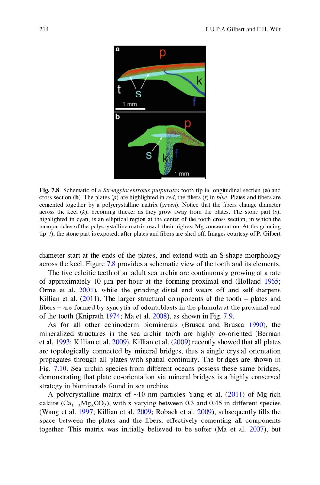
across the keel. Figure 7.8 provides a schematic view of the tooth and its elements.
The five calcitic teeth of an adult sea urchin are continuously growing at a rate
of approximately 10 mm per hour at the forming proximal end (Holland 1965;
Orme et al. 2001), while the grinding distal end wears off and self-sharpens
Killian et al. (2011). The larger structural components of the tooth – plates and
fibers – are formed by syncytia of odontoblasts in the plumula at the proximal end
of the tooth (Kniprath 1974; Ma et al. 2008), as shown in Fig. 7.9.
As for all other echinoderm biominerals (Brusca and Brusca 1990), the
mineralized structures in the sea urchin tooth are highly co-oriented (Berman
et al. 1993; Killian et al. 2009). Killian et al. (2009) recently showed that all plates
are topologically connected by mineral bridges, thus a single crystal orientation
propagates through all plates with spatial continuity. The bridges are shown in
Fig. 7.10. Sea urchin species from different oceans possess these same bridges,
demonstrating that plate co-orientation via mineral bridges is a highly conserved
strategy in biominerals found in sea urchins.
A polycrystalline matrix of ~10 nm particles Yang et al. (2011) of Mg-rich
calcite (Ca 1Àx Mg x CO 3 ), with x varying between 0.3 and 0.45 in different species
(Wang et al. 1997; Killian et al. 2009; Robach et al. 2009), subsequently fills the
space between the plates and the fibers, effectively cementing all components
together. This matrix was initially believed to be softer (Ma et al. 2007), but
Fig. 7.8 Schematic of a Strongylocentrotus purpuratus tooth tip in longitudinal section (a) and
cross section (b). The plates (p) are highlighted in red, the fibers (f) in blue. Plates and fibers are
cemented together by a polycrystalline matrix (green). Notice that the fibers change diameter
across the keel (k), becoming thicker as they grow away from the plates. The stone part (s),
highlighted in cyan, is an elliptical region at the center of the tooth cross section, in which the
nanoparticles of the polycrystalline matrix reach their highest Mg concentration. At the grinding
tip (t), the stone part is exposed, after plates and fibers are shed off. Images courtesy of P. Gilbert
214
P.U.P.A Gilbert and F.H. Wilt
across the keel. Figure 7.8 provides a schematic view of the tooth and its elements.
The five calcitic teeth of an adult sea urchin are continuously growing at a rate
of approximately 10 mm per hour at the forming proximal end (Holland 1965;
Orme et al. 2001), while the grinding distal end wears off and self-sharpens
Killian et al. (2011). The larger structural components of the tooth – plates and
fibers – are formed by syncytia of odontoblasts in the plumula at the proximal end
of the tooth (Kniprath 1974; Ma et al. 2008), as shown in Fig. 7.9.
As for all other echinoderm biominerals (Brusca and Brusca 1990), the
mineralized structures in the sea urchin tooth are highly co-oriented (Berman
et al. 1993; Killian et al. 2009). Killian et al. (2009) recently showed that all plates
are topologically connected by mineral bridges, thus a single crystal orientation
propagates through all plates with spatial continuity. The bridges are shown in
Fig. 7.10. Sea urchin species from different oceans possess these same bridges,
demonstrating that plate co-orientation via mineral bridges is a highly conserved
strategy in biominerals found in sea urchins.
A polycrystalline matrix of ~10 nm particles Yang et al. (2011) of Mg-rich
calcite (Ca 1Àx Mg x CO 3 ), with x varying between 0.3 and 0.45 in different species
(Wang et al. 1997; Killian et al. 2009; Robach et al. 2009), subsequently fills the
space between the plates and the fibers, effectively cementing all components
together. This matrix was initially believed to be softer (Ma et al. 2007), but
Fig. 7.8 Schematic of a Strongylocentrotus purpuratus tooth tip in longitudinal section (a) and
cross section (b). The plates (p) are highlighted in red, the fibers (f) in blue. Plates and fibers are
cemented together by a polycrystalline matrix (green). Notice that the fibers change diameter
across the keel (k), becoming thicker as they grow away from the plates. The stone part (s),
highlighted in cyan, is an elliptical region at the center of the tooth cross section, in which the
nanoparticles of the polycrystalline matrix reach their highest Mg concentration. At the grinding
tip (t), the stone part is exposed, after plates and fibers are shed off. Images courtesy of P. Gilbert
214
P.U.P.A Gilbert and F.H. Wilt