

et al. 1997; Wang 1998; Ma et al. 2007). The top part of the T is commonly termed
the flange; the vertical part of the T is the keel. The flange contains curved calcitic
plates, spaced at a few microns from each other. Calcitic fibers with varying
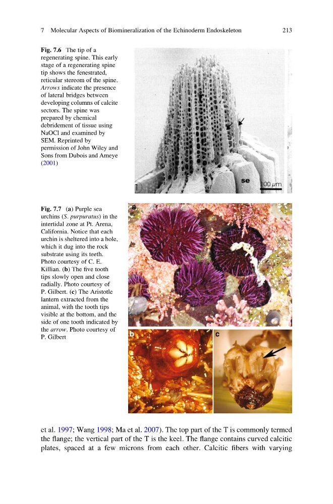
Fig. 7.7 (a) Purple sea
urchins (S. purpuratus) in the
intertidal zone at Pt. Arena,
California. Notice that each
urchin is sheltered into a hole,
which it dug into the rock
substrate using its teeth.
Photo courtesy of C. E.
Killian. (b) The five tooth
tips slowly open and close
radially. Photo courtesy of
P. Gilbert. (c) The Aristotle
lantern extracted from the
animal, with the tooth tips
visible at the bottom, and the
side of one tooth indicated by
the arrow. Photo courtesy of
P. Gilbert
Fig. 7.6 The tip of a
regenerating spine. This early
stage of a regenerating spine
tip shows the fenestrated,
reticular stereom of the spine.
Arrows indicate the presence
of lateral bridges between
developing columns of calcite
sectors. The spine was
prepared by chemical
debridement of tissue using
NaOCl and examined by
SEM. Reprinted by
permission of John Wiley and
Sons from Dubois and Ameye
(2001)
7 Molecular Aspects of Biomineralization of the Echinoderm Endoskeleton
213
the flange; the vertical part of the T is the keel. The flange contains curved calcitic
plates, spaced at a few microns from each other. Calcitic fibers with varying
Fig. 7.7 (a) Purple sea
urchins (S. purpuratus) in the
intertidal zone at Pt. Arena,
California. Notice that each
urchin is sheltered into a hole,
which it dug into the rock
substrate using its teeth.
Photo courtesy of C. E.
Killian. (b) The five tooth
tips slowly open and close
radially. Photo courtesy of
P. Gilbert. (c) The Aristotle
lantern extracted from the
animal, with the tooth tips
visible at the bottom, and the
side of one tooth indicated by
the arrow. Photo courtesy of
P. Gilbert
Fig. 7.6 The tip of a
regenerating spine. This early
stage of a regenerating spine
tip shows the fenestrated,
reticular stereom of the spine.
Arrows indicate the presence
of lateral bridges between
developing columns of calcite
sectors. The spine was
prepared by chemical
debridement of tissue using
NaOCl and examined by
SEM. Reprinted by
permission of John Wiley and
Sons from Dubois and Ameye
(2001)
7 Molecular Aspects of Biomineralization of the Echinoderm Endoskeleton
213