

and Banfield in TiO 2 (Penn and Banfield 1999), and FeOOH (Banfield et al. 2000),
and later in many other synthetic mesocrystals (C€ olfen and Antonietti 2008)? Or, is
this the result of an amorphous precursor phase forming first, with crystallinity
propagating through it subsequently (Aizenberg et al. 2003; Politi et al. 2008)?
This question was addressed in detail by Killian et al. (2009), who showed that,
indeed, amorphous precursor phases are aggregated first, and then crystallinity
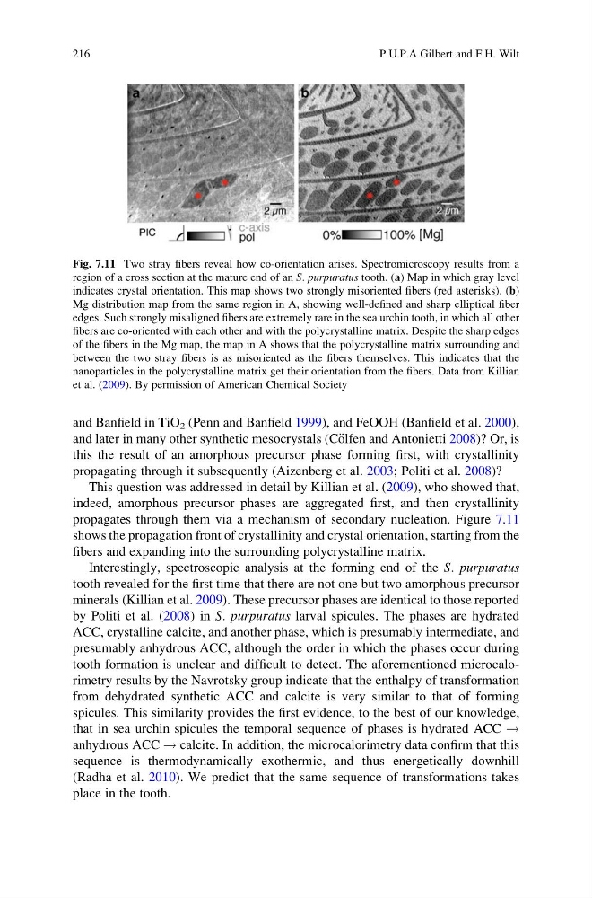
propagates through them via a mechanism of secondary nucleation. Figure 7.11
shows the propagation front of crystallinity and crystal orientation, starting from the
fibers and expanding into the surrounding polycrystalline matrix.
Interestingly, spectroscopic analysis at the forming end of the S. purpuratus
tooth revealed for the first time that there are not one but two amorphous precursor
minerals (Killian et al. 2009). These precursor phases are identical to those reported
by Politi et al. (2008) in S. purpuratus larval spicules. The phases are hydrated
ACC, crystalline calcite, and another phase, which is presumably intermediate, and
presumably anhydrous ACC, although the order in which the phases occur during
tooth formation is unclear and difficult to detect. The aforementioned microcalorimetry results by the Navrotsky group indicate that the enthalpy of transformation
from dehydrated synthetic ACC and calcite is very similar to that of forming
spicules. This similarity provides the first evidence, to the best of our knowledge,
that in sea urchin spicules the temporal sequence of phases is hydrated ACC !
anhydrous ACC ! calcite. In addition, the microcalorimetry data confirm that this
sequence is thermodynamically exothermic, and thus energetically downhill
(Radha et al. 2010). We predict that the same sequence of transformations takes
place in the tooth.
Fig. 7.11 Two stray fibers reveal how co-orientation arises. Spectromicroscopy results from a
region of a cross section at the mature end of an S. purpuratus tooth. (a) Map in which gray level
indicates crystal orientation. This map shows two strongly misoriented fibers (red asterisks). (b)
Mg distribution map from the same region in A, showing well-defined and sharp elliptical fiber
edges. Such strongly misaligned fibers are extremely rare in the sea urchin tooth, in which all other
fibers are co-oriented with each other and with the polycrystalline matrix. Despite the sharp edges
of the fibers in the Mg map, the map in A shows that the polycrystalline matrix surrounding and
between the two stray fibers is as misoriented as the fibers themselves. This indicates that the
nanoparticles in the polycrystalline matrix get their orientation from the fibers. Data from Killian
et al. (2009). By permission of American Chemical Society
216
P.U.P.A Gilbert and F.H. Wilt
and later in many other synthetic mesocrystals (C€ olfen and Antonietti 2008)? Or, is
this the result of an amorphous precursor phase forming first, with crystallinity
propagating through it subsequently (Aizenberg et al. 2003; Politi et al. 2008)?
This question was addressed in detail by Killian et al. (2009), who showed that,
indeed, amorphous precursor phases are aggregated first, and then crystallinity
propagates through them via a mechanism of secondary nucleation. Figure 7.11
shows the propagation front of crystallinity and crystal orientation, starting from the
fibers and expanding into the surrounding polycrystalline matrix.
Interestingly, spectroscopic analysis at the forming end of the S. purpuratus
tooth revealed for the first time that there are not one but two amorphous precursor
minerals (Killian et al. 2009). These precursor phases are identical to those reported
by Politi et al. (2008) in S. purpuratus larval spicules. The phases are hydrated
ACC, crystalline calcite, and another phase, which is presumably intermediate, and
presumably anhydrous ACC, although the order in which the phases occur during
tooth formation is unclear and difficult to detect. The aforementioned microcalorimetry results by the Navrotsky group indicate that the enthalpy of transformation
from dehydrated synthetic ACC and calcite is very similar to that of forming
spicules. This similarity provides the first evidence, to the best of our knowledge,
that in sea urchin spicules the temporal sequence of phases is hydrated ACC !
anhydrous ACC ! calcite. In addition, the microcalorimetry data confirm that this
sequence is thermodynamically exothermic, and thus energetically downhill
(Radha et al. 2010). We predict that the same sequence of transformations takes
place in the tooth.
Fig. 7.11 Two stray fibers reveal how co-orientation arises. Spectromicroscopy results from a
region of a cross section at the mature end of an S. purpuratus tooth. (a) Map in which gray level
indicates crystal orientation. This map shows two strongly misoriented fibers (red asterisks). (b)
Mg distribution map from the same region in A, showing well-defined and sharp elliptical fiber
edges. Such strongly misaligned fibers are extremely rare in the sea urchin tooth, in which all other
fibers are co-oriented with each other and with the polycrystalline matrix. Despite the sharp edges
of the fibers in the Mg map, the map in A shows that the polycrystalline matrix surrounding and
between the two stray fibers is as misoriented as the fibers themselves. This indicates that the
nanoparticles in the polycrystalline matrix get their orientation from the fibers. Data from Killian
et al. (2009). By permission of American Chemical Society
216
P.U.P.A Gilbert and F.H. Wilt