

using microincrements as records of fishes’ daily growth. The existence of daily
increments in otolith is believed to be a widespread phenomenon among fish
according to Campana and Neilson (1985). They considered that the otolith was
composed of an incremental zone and a discontinuous zone. The former zone is
wide and transparent, composed of calcium carbonate. The latter zone is narrow and
opaque, composed of organic matrix. Current research works are mainly restricted
to the field of the daily ring structure. Understanding microstructure of otolith is
important for studying the larvae and juvenile fish. Campana and Neilson (1985)
studied the ecological effect on otolith’s microstructure, including the early life
history, age, growth, recruitment, migration, mortality, stock structure, and recording the historical life information. Otolith microstructure examination and analysis
could be used to discriminate populations, to show the feeding and growth history
of fish experienced in wild or reared conditions in the elapsed days. The microstructure of otolith is related to many factors. First of all, the environmental
conditions, such as water temperature, water enrichment elements, food supply,
etc., could influence the increment width, contrast, transparence, and morphology.
The otolith’s growth and structure are also affected by proteins. Sollner et al.
reported a gene, starmaker, which can control the crystal lattice structure and the
shape of the otolith (Sollner et al. 2003).
Calcitic Microlenses in Brittlestar
Photosensitivity in most echinoderms has been attributed to “diffuse” dermal
receptors. Aizenberg et al. (2001) reported that certain single calcite crystals used
by brittlestars for skeletal construction are also a component of specialized
photosensory organs, conceivably with the function of a compound eye. The
analysis of arm ossicles in Ophiocoma showed that in light-sensitive species, the
periphery of the labyrinthic calcitic skeleton extends into a regular array of spherical microstructures that have a characteristic double-lens design. These structures
are absent in light-indifferent species. Photolithographic experiments in which a
photoresist film was illuminated through the lens array showed selective exposure
of the photoresist under the lens centers. These results provide experimental
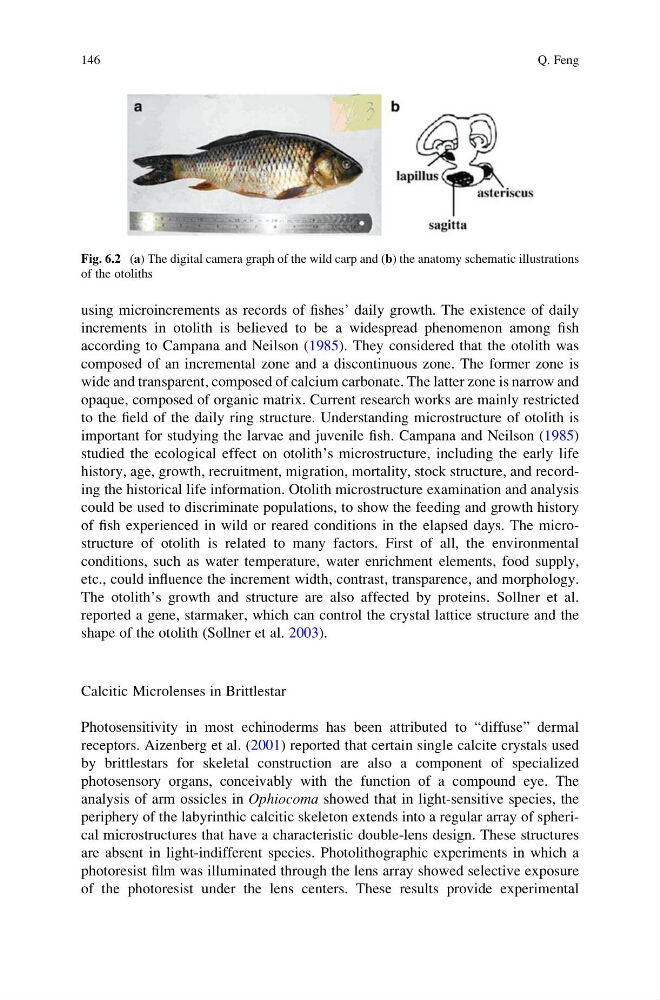
Fig. 6.2 (a) The digital camera graph of the wild carp and (b) the anatomy schematic illustrations
of the otoliths
146
Q. Feng
increments in otolith is believed to be a widespread phenomenon among fish
according to Campana and Neilson (1985). They considered that the otolith was
composed of an incremental zone and a discontinuous zone. The former zone is
wide and transparent, composed of calcium carbonate. The latter zone is narrow and
opaque, composed of organic matrix. Current research works are mainly restricted
to the field of the daily ring structure. Understanding microstructure of otolith is
important for studying the larvae and juvenile fish. Campana and Neilson (1985)
studied the ecological effect on otolith’s microstructure, including the early life
history, age, growth, recruitment, migration, mortality, stock structure, and recording the historical life information. Otolith microstructure examination and analysis
could be used to discriminate populations, to show the feeding and growth history
of fish experienced in wild or reared conditions in the elapsed days. The microstructure of otolith is related to many factors. First of all, the environmental
conditions, such as water temperature, water enrichment elements, food supply,
etc., could influence the increment width, contrast, transparence, and morphology.
The otolith’s growth and structure are also affected by proteins. Sollner et al.
reported a gene, starmaker, which can control the crystal lattice structure and the
shape of the otolith (Sollner et al. 2003).
Calcitic Microlenses in Brittlestar
Photosensitivity in most echinoderms has been attributed to “diffuse” dermal
receptors. Aizenberg et al. (2001) reported that certain single calcite crystals used
by brittlestars for skeletal construction are also a component of specialized
photosensory organs, conceivably with the function of a compound eye. The
analysis of arm ossicles in Ophiocoma showed that in light-sensitive species, the
periphery of the labyrinthic calcitic skeleton extends into a regular array of spherical microstructures that have a characteristic double-lens design. These structures
are absent in light-indifferent species. Photolithographic experiments in which a
photoresist film was illuminated through the lens array showed selective exposure
of the photoresist under the lens centers. These results provide experimental
Fig. 6.2 (a) The digital camera graph of the wild carp and (b) the anatomy schematic illustrations
of the otoliths
146
Q. Feng