

evidence that the microlenses are optical elements that guide and focus the light
inside the tissue. The estimated focal distance (4–7 mm below the lenses) coincides
with the location of nerve bundles, the presumed primary photoreceptors. The lens
array is designed to minimize spherical aberration and birefringence and to detect
light from a particular direction. The optical performance is further optimized by
phototropic chromatophores that regulate the dose of illumination reaching the
receptors. These structures represent an example of a multifunctional biomaterial
that fulfills both mechanical and optical functions.
Echinoderms in general, and especially the brittlestars (Ophiuroidea), exhibit a
wide range of responses to light intensity, from a largely light-indifferent behavior
to pronounced color change and rapid escape behavior (Aizenberg et al. 2001).
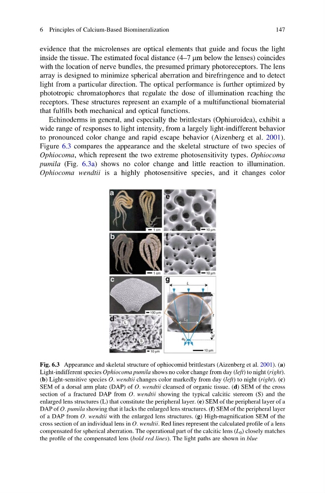
Figure 6.3 compares the appearance and the skeletal structure of two species of
Ophiocoma, which represent the two extreme photosensitivity types. Ophiocoma
pumila (Fig. 6.3a) shows no color change and little reaction to illumination.
Ophiocoma wendtii is a highly photosensitive species, and it changes color
Fig. 6.3 Appearance and skeletal structure of ophiocomid brittlestars (Aizenberg et al. 2001). (a)
Light-indifferent species Ophiocoma pumila shows no color change from day (left) to night (right).
(b) Light-sensitive species O. wendtii changes color markedly from day (left) to night (right). (c)
SEM of a dorsal arm plate (DAP) of O. wendtii cleansed of organic tissue. (d) SEM of the cross
section of a fractured DAP from O. wendtii showing the typical calcitic stereom (S) and the
enlarged lens structures (L) that constitute the peripheral layer. (e) SEM of the peripheral layer of a
DAP of O. pumila showing that it lacks the enlarged lens structures. (f) SEM of the peripheral layer
of a DAP from O. wendtii with the enlarged lens structures. (g) High-magnification SEM of the
cross section of an individual lens in O. wendtii. Red lines represent the calculated profile of a lens
compensated for spherical aberration. The operational part of the calcitic lens (L 0 ) closely matches
the profile of the compensated lens (bold red lines). The light paths are shown in blue
6 Principles of Calcium-Based Biomineralization
147
inside the tissue. The estimated focal distance (4–7 mm below the lenses) coincides
with the location of nerve bundles, the presumed primary photoreceptors. The lens
array is designed to minimize spherical aberration and birefringence and to detect
light from a particular direction. The optical performance is further optimized by
phototropic chromatophores that regulate the dose of illumination reaching the
receptors. These structures represent an example of a multifunctional biomaterial
that fulfills both mechanical and optical functions.
Echinoderms in general, and especially the brittlestars (Ophiuroidea), exhibit a
wide range of responses to light intensity, from a largely light-indifferent behavior
to pronounced color change and rapid escape behavior (Aizenberg et al. 2001).
Figure 6.3 compares the appearance and the skeletal structure of two species of
Ophiocoma, which represent the two extreme photosensitivity types. Ophiocoma
pumila (Fig. 6.3a) shows no color change and little reaction to illumination.
Ophiocoma wendtii is a highly photosensitive species, and it changes color
Fig. 6.3 Appearance and skeletal structure of ophiocomid brittlestars (Aizenberg et al. 2001). (a)
Light-indifferent species Ophiocoma pumila shows no color change from day (left) to night (right).
(b) Light-sensitive species O. wendtii changes color markedly from day (left) to night (right). (c)
SEM of a dorsal arm plate (DAP) of O. wendtii cleansed of organic tissue. (d) SEM of the cross
section of a fractured DAP from O. wendtii showing the typical calcitic stereom (S) and the
enlarged lens structures (L) that constitute the peripheral layer. (e) SEM of the peripheral layer of a
DAP of O. pumila showing that it lacks the enlarged lens structures. (f) SEM of the peripheral layer
of a DAP from O. wendtii with the enlarged lens structures. (g) High-magnification SEM of the
cross section of an individual lens in O. wendtii. Red lines represent the calculated profile of a lens
compensated for spherical aberration. The operational part of the calcitic lens (L 0 ) closely matches
the profile of the compensated lens (bold red lines). The light paths are shown in blue
6 Principles of Calcium-Based Biomineralization
147