

4.5.5 Elemental Mapping
Elemental distribution (mapping) of the crust samples was qualitatively assessed by
an energy-dispersive X-ray spectrometer coupled to a focused ion beam (FIB)assisted SEM (Wang et al. 2009a).
For elemental mapping, we have selected one coccolith and its nearest surrounding to determine the spatial distribution of the elements within a coccolith
fragment. For this study, a representative endothecal coccolith, existing within a
coccosphere, was selected (Fig. 4.16a); this coccolith was well structured and
displayed in the central area the characteristic openings (Fig. 4.16b). Elemental
maps of the Ka-lines for Ca (Fig. 165c), Mn (Fig. 4.16d), Si (Fig. 4.16e), O, and Na
were recorded using different acquisition channels. The advantage of this technique
is the colocalization of the elements with the scanning electronic image. It is
evident from the elemental maps that high signals, matching the structure of the
coccolith, are seen for Ca and Mn (Fig. 4.16c and d). In contrast, the intensities for
Si (Fig. 4.16e), O, and Na were lower at the coccolith structure, compared to the
surrounding structures. This series of experiments showed that a high accumulation
of both Ca together with Mn exists in the coccoliths, while no Fe can be traced there
(to be published). This finding provided the clue to the participation of coccoliths as
bio-seeds during Mn precipitation in crusts.
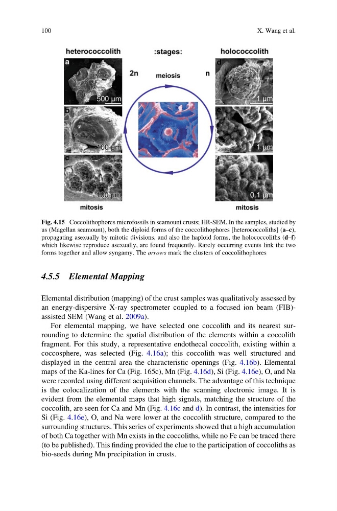
Fig. 4.15 Coccolithophores microfossils in seamount crusts; HR-SEM. In the samples, studied by
us (Magellan seamount), both the diploid forms of the coccolithophores [heterococcoliths] (a–c),
propagating asexually by mitotic divisions, and also the haploid forms, the holococcoliths (d–f)
which likewise reproduce asexually, are found frequently. Rarely occurring events link the two
forms together and allow syngamy. The arrows mark the clusters of coccolithophores
100
X. Wang et al.
Elemental distribution (mapping) of the crust samples was qualitatively assessed by
an energy-dispersive X-ray spectrometer coupled to a focused ion beam (FIB)assisted SEM (Wang et al. 2009a).
For elemental mapping, we have selected one coccolith and its nearest surrounding to determine the spatial distribution of the elements within a coccolith
fragment. For this study, a representative endothecal coccolith, existing within a
coccosphere, was selected (Fig. 4.16a); this coccolith was well structured and
displayed in the central area the characteristic openings (Fig. 4.16b). Elemental
maps of the Ka-lines for Ca (Fig. 165c), Mn (Fig. 4.16d), Si (Fig. 4.16e), O, and Na
were recorded using different acquisition channels. The advantage of this technique
is the colocalization of the elements with the scanning electronic image. It is
evident from the elemental maps that high signals, matching the structure of the
coccolith, are seen for Ca and Mn (Fig. 4.16c and d). In contrast, the intensities for
Si (Fig. 4.16e), O, and Na were lower at the coccolith structure, compared to the
surrounding structures. This series of experiments showed that a high accumulation
of both Ca together with Mn exists in the coccoliths, while no Fe can be traced there
(to be published). This finding provided the clue to the participation of coccoliths as
bio-seeds during Mn precipitation in crusts.
Fig. 4.15 Coccolithophores microfossils in seamount crusts; HR-SEM. In the samples, studied by
us (Magellan seamount), both the diploid forms of the coccolithophores [heterococcoliths] (a–c),
propagating asexually by mitotic divisions, and also the haploid forms, the holococcoliths (d–f)
which likewise reproduce asexually, are found frequently. Rarely occurring events link the two
forms together and allow syngamy. The arrows mark the clusters of coccolithophores
100
X. Wang et al.