

4.4.4 Bio-seedS
Solid ground for the involvement of bacteria in polymetallic nodule formation had
been presented by Ehrlich (see: Ehrlich 2002). However, only recently was it possible
to visualize distinct bacteria by in situ HR-SEM analysis within the mineralized
deposits of the nodules, and not on their surfaces (Wang and M€ uller 2009; Wang
et al. 2009b, c). Analysis of small, broken splinters revealed two bacterial
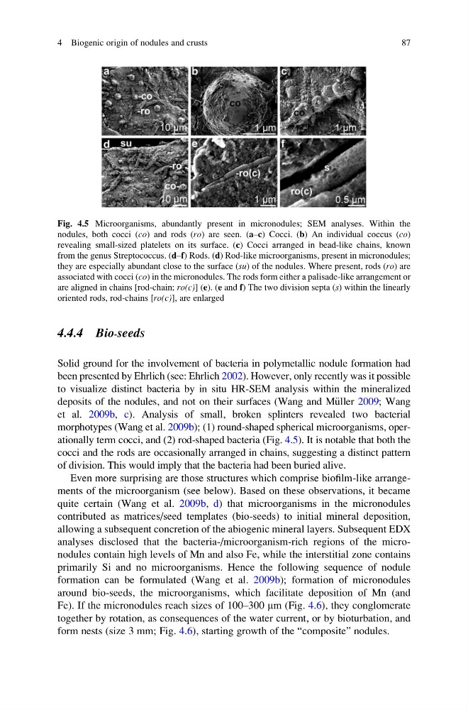
morphotypes (Wang et al. 2009b); (1) round-shaped spherical microorganisms, operationally term cocci, and (2) rod-shaped bacteria (Fig. 4.5). It is notable that both the
cocci and the rods are occasionally arranged in chains, suggesting a distinct pattern
of division. This would imply that the bacteria had been buried alive.
Even more surprising are those structures which comprise biofilm-like arrangements of the microorganism (see below). Based on these observations, it became
quite certain (Wang et al. 2009b, d) that microorganisms in the micronodules
contributed as matrices/seed templates (bio-seeds) to initial mineral deposition,
allowing a subsequent concretion of the abiogenic mineral layers. Subsequent EDX
analyses disclosed that the bacteria-/microorganism-rich regions of the micronodules contain high levels of Mn and also Fe, while the interstitial zone contains
primarily Si and no microorganisms. Hence the following sequence of nodule
formation can be formulated (Wang et al. 2009b); formation of micronodules
around bio-seeds, the microorganisms, which facilitate deposition of Mn (and
Fe). If the micronodules reach sizes of 100–300 mm (Fig. 4.6), they conglomerate
together by rotation, as consequences of the water current, or by bioturbation, and
form nests (size 3 mm; Fig. 4.6), starting growth of the “composite” nodules.
Fig. 4.5 Microorganisms, abundantly present in micronodules; SEM analyses. Within the
nodules, both cocci (co) and rods (ro) are seen. (a–c) Cocci. (b) An individual coccus (co)
revealing small-sized platelets on its surface. (c) Cocci arranged in bead-like chains, known
from the genus Streptococcus. (d–f) Rods. (d) Rod-like microorganisms, present in micronodules;
they are especially abundant close to the surface (su) of the nodules. Where present, rods (ro) are
associated with cocci (co) in the micronodules. The rods form either a palisade-like arrangement or
are aligned in chains [rod-chain; ro(c)] (e). (e and f) The two division septa (s) within the linearly
oriented rods, rod-chains [ro(c)], are enlarged
4 Biogenic origin of nodules and crusts
87
Solid ground for the involvement of bacteria in polymetallic nodule formation had
been presented by Ehrlich (see: Ehrlich 2002). However, only recently was it possible
to visualize distinct bacteria by in situ HR-SEM analysis within the mineralized
deposits of the nodules, and not on their surfaces (Wang and M€ uller 2009; Wang
et al. 2009b, c). Analysis of small, broken splinters revealed two bacterial
morphotypes (Wang et al. 2009b); (1) round-shaped spherical microorganisms, operationally term cocci, and (2) rod-shaped bacteria (Fig. 4.5). It is notable that both the
cocci and the rods are occasionally arranged in chains, suggesting a distinct pattern
of division. This would imply that the bacteria had been buried alive.
Even more surprising are those structures which comprise biofilm-like arrangements of the microorganism (see below). Based on these observations, it became
quite certain (Wang et al. 2009b, d) that microorganisms in the micronodules
contributed as matrices/seed templates (bio-seeds) to initial mineral deposition,
allowing a subsequent concretion of the abiogenic mineral layers. Subsequent EDX
analyses disclosed that the bacteria-/microorganism-rich regions of the micronodules contain high levels of Mn and also Fe, while the interstitial zone contains
primarily Si and no microorganisms. Hence the following sequence of nodule
formation can be formulated (Wang et al. 2009b); formation of micronodules
around bio-seeds, the microorganisms, which facilitate deposition of Mn (and
Fe). If the micronodules reach sizes of 100–300 mm (Fig. 4.6), they conglomerate
together by rotation, as consequences of the water current, or by bioturbation, and
form nests (size 3 mm; Fig. 4.6), starting growth of the “composite” nodules.
Fig. 4.5 Microorganisms, abundantly present in micronodules; SEM analyses. Within the
nodules, both cocci (co) and rods (ro) are seen. (a–c) Cocci. (b) An individual coccus (co)
revealing small-sized platelets on its surface. (c) Cocci arranged in bead-like chains, known
from the genus Streptococcus. (d–f) Rods. (d) Rod-like microorganisms, present in micronodules;
they are especially abundant close to the surface (su) of the nodules. Where present, rods (ro) are
associated with cocci (co) in the micronodules. The rods form either a palisade-like arrangement or
are aligned in chains [rod-chain; ro(c)] (e). (e and f) The two division septa (s) within the linearly
oriented rods, rod-chains [ro(c)], are enlarged
4 Biogenic origin of nodules and crusts
87