

each other at their longitudinal surfaces, or (2) linearly arranged as it is often seen in
the taxa of Eubacteria (Fig. 4.5e and f). The rods were about 1.5–2.5-mm long and
0.35–0.45-mm thick (Fig. 4.5d and e). From the microscopic images, it appeared
that the rods divided medially (Szeto et al. 2001). Such a division pattern is known
from bacilli [Streptobacilli (Ryan and Ray 2004)]: The rods appear in chains; a
chain with three attached rods is shown in Fig. 4.5e and f, together with the two
division septa. It remains unsolved if the rods separated completely after division –
which is most likely – or remained fused. The surrounding, adjacent zones of the
rods were formed by solid material, which appeared homogenous.
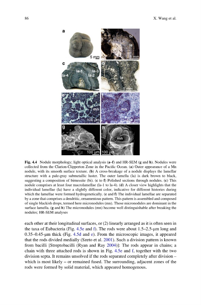
Fig. 4.4 Nodule morphology; light optical analysis (a–f) and HR-SEM (g and h). Nodules were
collected from the Clarion-Clipperton Zone in the Pacific Ocean. (a) Outer appearance of a Mn
nodule, with its smooth surface texture. (b) A cross-breakage of a nodule displays the lamellar
structure with a pale-gray submetallic luster. The outer lamella (la) is dark brown to black,
suggesting a composition of birnessite (bi). (c to f) Polished sections through nodules. (c) This
nodule comprises at least four macrolamellae (la-1 to la-4). (d) A closer view highlights that the
individual lamellae (la) have a slightly different color, indicative for different histories during
which the lamellae were formed hydrogenetically. (e and f) The individual lamellae are separated
by a zone that comprises a dendritic, ornamentous pattern. This pattern is assembled and composed
of single blackish drops, termed here micronodules (mn). Those micronodules are dominant in the
surface lamella. (g and h) The micronodules (mn) become well distinguishable after breaking the
nodules; HR-SEM analyses
86
X. Wang et al.
the taxa of Eubacteria (Fig. 4.5e and f). The rods were about 1.5–2.5-mm long and
0.35–0.45-mm thick (Fig. 4.5d and e). From the microscopic images, it appeared
that the rods divided medially (Szeto et al. 2001). Such a division pattern is known
from bacilli [Streptobacilli (Ryan and Ray 2004)]: The rods appear in chains; a
chain with three attached rods is shown in Fig. 4.5e and f, together with the two
division septa. It remains unsolved if the rods separated completely after division –
which is most likely – or remained fused. The surrounding, adjacent zones of the
rods were formed by solid material, which appeared homogenous.
Fig. 4.4 Nodule morphology; light optical analysis (a–f) and HR-SEM (g and h). Nodules were
collected from the Clarion-Clipperton Zone in the Pacific Ocean. (a) Outer appearance of a Mn
nodule, with its smooth surface texture. (b) A cross-breakage of a nodule displays the lamellar
structure with a pale-gray submetallic luster. The outer lamella (la) is dark brown to black,
suggesting a composition of birnessite (bi). (c to f) Polished sections through nodules. (c) This
nodule comprises at least four macrolamellae (la-1 to la-4). (d) A closer view highlights that the
individual lamellae (la) have a slightly different color, indicative for different histories during
which the lamellae were formed hydrogenetically. (e and f) The individual lamellae are separated
by a zone that comprises a dendritic, ornamentous pattern. This pattern is assembled and composed
of single blackish drops, termed here micronodules (mn). Those micronodules are dominant in the
surface lamella. (g and h) The micronodules (mn) become well distinguishable after breaking the
nodules; HR-SEM analyses
86
X. Wang et al.