

Microfluidic Systems for Marine Biotechnology 20.3 Microfluidic Devices for Sea-Related Health and Safety 519
Part C | 20.3
a)
b)
Fluidic layer
Buffer circle
Sample circle
Regular valve
Sieve valve
Reaction column
Cell solution
Buffer inlet 1
Buffer inlet 2
Microcages
Outlet
Sample
inlet
Syringe pumps
Microfluidic
chip
Buffer
solution
Ultrasonication
bath
Outlet
reservoir
Piezoelectric element
Control layer
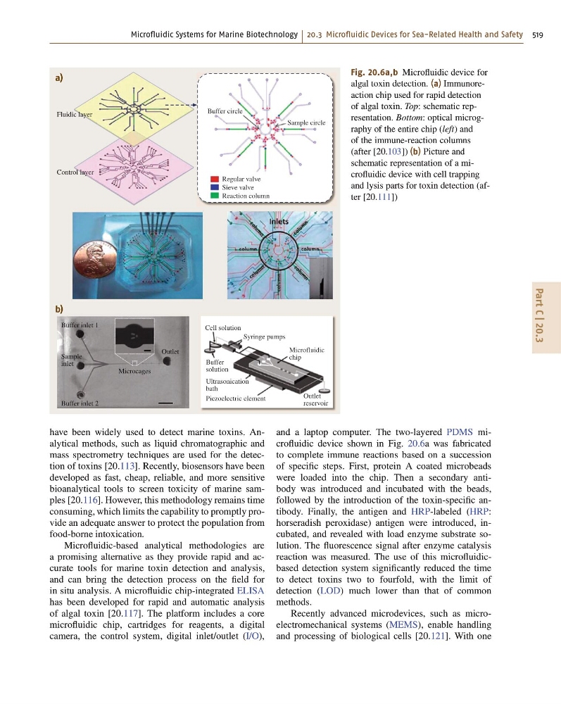
Fig. 20.6a,b Microfluidic device for
algal toxin detection. (a) Immunoreaction chip used for rapid detection
of algal toxin. Top: schematic representation. Bottom: optical micrography of the entire chip (left) and
of the immune-reaction columns
(after [20.103]) (b) Picture and
schematic representation of a microfluidic device with cell trapping
and lysis parts for toxin detection (after [20.111])
have been widely used to detect marine toxins. Analytical methods, such as liquid chromatographic and
mass spectrometry techniques are used for the detection of toxins [20.113]. Recently, biosensors have been
developed as fast, cheap, reliable, and more sensitive
bioanalytical tools to screen toxicity of marine samples [20.116]. However, this methodology remains time
consuming, which limits the capability to promptly provide an adequate answer to protect the population from
food-borne intoxication.
Microfluidic-based analytical methodologies are
a promising alternative as they provide rapid and accurate tools for marine toxin detection and analysis,
and can bring the detection process on the field for
in situ analysis. A microfluidic chip-integrated ELISA
has been developed for rapid and automatic analysis
of algal toxin [20.117]. The platform includes a core
microfluidic chip, cartridges for reagents, a digital
camera, the control system, digital inlet/outlet (I/O),
and a laptop computer. The two-layered PDMS microfluidic device shown in Fig. 20.6a was fabricated
to complete immune reactions based on a succession
of specific steps. First, protein A coated microbeads
were loaded into the chip. Then a secondary antibody was introduced and incubated with the beads,
followed by the introduction of the toxin-specific antibody. Finally, the antigen and HRP-labeled (HRP:
horseradish peroxidase) antigen were introduced, incubated, and revealed with load enzyme substrate solution. The fluorescence signal after enzyme catalysis
reaction was measured. The use of this microfluidicbased detection system significantly reduced the time
to detect toxins two to fourfold, with the limit of
detection (LOD) much lower than that of common
methods.
Recently advanced microdevices, such as microelectromechanical systems (MEMS), enable handling
and processing of biological cells [20.121]. With one
Part C | 20.3
a)
b)
Fluidic layer
Buffer circle
Sample circle
Regular valve
Sieve valve
Reaction column
Cell solution
Buffer inlet 1
Buffer inlet 2
Microcages
Outlet
Sample
inlet
Syringe pumps
Microfluidic
chip
Buffer
solution
Ultrasonication
bath
Outlet
reservoir
Piezoelectric element
Control layer
Fig. 20.6a,b Microfluidic device for
algal toxin detection. (a) Immunoreaction chip used for rapid detection
of algal toxin. Top: schematic representation. Bottom: optical micrography of the entire chip (left) and
of the immune-reaction columns
(after [20.103]) (b) Picture and
schematic representation of a microfluidic device with cell trapping
and lysis parts for toxin detection (after [20.111])
have been widely used to detect marine toxins. Analytical methods, such as liquid chromatographic and
mass spectrometry techniques are used for the detection of toxins [20.113]. Recently, biosensors have been
developed as fast, cheap, reliable, and more sensitive
bioanalytical tools to screen toxicity of marine samples [20.116]. However, this methodology remains time
consuming, which limits the capability to promptly provide an adequate answer to protect the population from
food-borne intoxication.
Microfluidic-based analytical methodologies are
a promising alternative as they provide rapid and accurate tools for marine toxin detection and analysis,
and can bring the detection process on the field for
in situ analysis. A microfluidic chip-integrated ELISA
has been developed for rapid and automatic analysis
of algal toxin [20.117]. The platform includes a core
microfluidic chip, cartridges for reagents, a digital
camera, the control system, digital inlet/outlet (I/O),
and a laptop computer. The two-layered PDMS microfluidic device shown in Fig. 20.6a was fabricated
to complete immune reactions based on a succession
of specific steps. First, protein A coated microbeads
were loaded into the chip. Then a secondary antibody was introduced and incubated with the beads,
followed by the introduction of the toxin-specific antibody. Finally, the antigen and HRP-labeled (HRP:
horseradish peroxidase) antigen were introduced, incubated, and revealed with load enzyme substrate solution. The fluorescence signal after enzyme catalysis
reaction was measured. The use of this microfluidicbased detection system significantly reduced the time
to detect toxins two to fourfold, with the limit of
detection (LOD) much lower than that of common
methods.
Recently advanced microdevices, such as microelectromechanical systems (MEMS), enable handling
and processing of biological cells [20.121]. With one