

Part C | 20.3
518 Part C Marine Genomics
a)
c)
d)
b)
Microchannel
(100 µm ×100 µm)
Curved
waveguide
(8 µm × 8µm)
Channel inlet
Differential
amplifier
SR844
RF lock-in amplifiers
Mixer
Frequency 2
Frequency 1
Objective
Flow
Rb
Rb
X1 Y1
X2 Y2
Vdiff
Channel
outlet
Algae
4-quad
photodetector
Itotal = A+B+C+D
ΔX = (A+B) – (C+D)
1
0
–1
–2
2.13
2.08
2.03
575 nm filter
532 nm filter
54 mm
85.6 mm
Beam dump
Sample
Sheath
660 nm filter
ΔX signal
Itotal signal
0
2 0
4 0
Time (ms)
Itotal
signal (V)
ΔX signal (V)
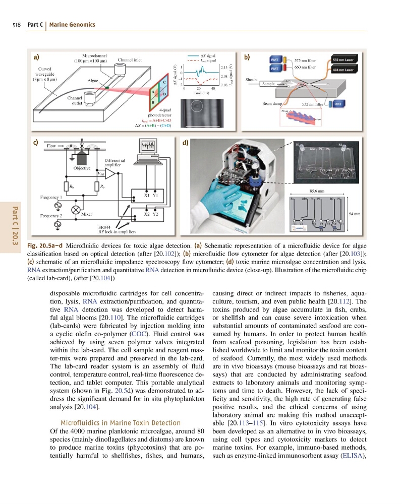
Fig. 20.5a–d Microfluidic devices for toxic algae detection. (a) Schematic representation of a microfluidic device for algae
classification based on optical detection (after [20.102]); (b) microfluidic flow cytometer for algae detection (after [20.103]);
(c) schematic of an microfluidic impedance spectroscopy flow cytometer; (d) toxic marine microalgae concentration and lysis,
RNA extraction/purification and quantitative RNA detection in microfluidic device (close-up). Illustration of the microfluidic chip
(called lab-card), (after [20.104])
disposable microfluidic cartridges for cell concentration, lysis, RNA extraction/purification, and quantitative RNA detection was developed to detect harmful algal blooms [20.110]. The microfluidic cartridges
(lab-cards) were fabricated by injection molding into
a cyclic olefin co-polymer (COC). Fluid control was
achieved by using seven polymer valves integrated
within the lab-card. The cell sample and reagent master-mix were prepared and preserved in the lab-card.
The lab-card reader system is an assembly of fluid
control, temperature control, real-time fluorescence detection, and tablet computer. This portable analytical
system (shown in Fig. 20.5d) was demonstrated to address the significant demand for in situ phytoplankton
analysis [20.104].
Microfluidics in Marine Toxin Detection
Of the 4000 marine planktonic microalgae, around 80
species (mainly dinoflagellates and diatoms) are known
to produce marine toxins (phycotoxins) that are potentially harmful to shellfishes, fishes, and humans,
causing direct or indirect impacts to fisheries, aquaculture, tourism, and even public health [20.112]. The
toxins produced by algae accumulate in fish, crabs,
or shellfish and can cause severe intoxication when
substantial amounts of contaminated seafood are consumed by humans. In order to protect human health
from seafood poisoning, legislation has been established worldwide to limit and monitor the toxin content
of seafood. Currently, the most widely used methods
are in vivo bioassays (mouse bioassays and rat bioassays) that are conducted by administrating seafood
extracts to laboratory animals and monitoring symptoms and time to death. However, the lack of specificity and sensitivity, the high rate of generating false
positive results, and the ethical concerns of using
laboratory animal are making this method unacceptable [20.113–115]. In vitro cytotoxicity assays have
been developed as an alternative to in vivo bioassays,
using cell types and cytotoxicity markers to detect
marine toxins. For example, immuno-based methods,
such as enzyme-linked immunosorbent assay (ELISA),
518 Part C Marine Genomics
a)
c)
d)
b)
Microchannel
(100 µm ×100 µm)
Curved
waveguide
(8 µm × 8µm)
Channel inlet
Differential
amplifier
SR844
RF lock-in amplifiers
Mixer
Frequency 2
Frequency 1
Objective
Flow
Rb
Rb
X1 Y1
X2 Y2
Vdiff
Channel
outlet
Algae
4-quad
photodetector
Itotal = A+B+C+D
ΔX = (A+B) – (C+D)
1
0
–1
–2
2.13
2.08
2.03
575 nm filter
532 nm filter
54 mm
85.6 mm
Beam dump
Sample
Sheath
660 nm filter
ΔX signal
Itotal signal
0
2 0
4 0
Time (ms)
Itotal
signal (V)
ΔX signal (V)
Fig. 20.5a–d Microfluidic devices for toxic algae detection. (a) Schematic representation of a microfluidic device for algae
classification based on optical detection (after [20.102]); (b) microfluidic flow cytometer for algae detection (after [20.103]);
(c) schematic of an microfluidic impedance spectroscopy flow cytometer; (d) toxic marine microalgae concentration and lysis,
RNA extraction/purification and quantitative RNA detection in microfluidic device (close-up). Illustration of the microfluidic chip
(called lab-card), (after [20.104])
disposable microfluidic cartridges for cell concentration, lysis, RNA extraction/purification, and quantitative RNA detection was developed to detect harmful algal blooms [20.110]. The microfluidic cartridges
(lab-cards) were fabricated by injection molding into
a cyclic olefin co-polymer (COC). Fluid control was
achieved by using seven polymer valves integrated
within the lab-card. The cell sample and reagent master-mix were prepared and preserved in the lab-card.
The lab-card reader system is an assembly of fluid
control, temperature control, real-time fluorescence detection, and tablet computer. This portable analytical
system (shown in Fig. 20.5d) was demonstrated to address the significant demand for in situ phytoplankton
analysis [20.104].
Microfluidics in Marine Toxin Detection
Of the 4000 marine planktonic microalgae, around 80
species (mainly dinoflagellates and diatoms) are known
to produce marine toxins (phycotoxins) that are potentially harmful to shellfishes, fishes, and humans,
causing direct or indirect impacts to fisheries, aquaculture, tourism, and even public health [20.112]. The
toxins produced by algae accumulate in fish, crabs,
or shellfish and can cause severe intoxication when
substantial amounts of contaminated seafood are consumed by humans. In order to protect human health
from seafood poisoning, legislation has been established worldwide to limit and monitor the toxin content
of seafood. Currently, the most widely used methods
are in vivo bioassays (mouse bioassays and rat bioassays) that are conducted by administrating seafood
extracts to laboratory animals and monitoring symptoms and time to death. However, the lack of specificity and sensitivity, the high rate of generating false
positive results, and the ethical concerns of using
laboratory animal are making this method unacceptable [20.113–115]. In vitro cytotoxicity assays have
been developed as an alternative to in vivo bioassays,
using cell types and cytotoxicity markers to detect
marine toxins. For example, immuno-based methods,
such as enzyme-linked immunosorbent assay (ELISA),