

Part C | 20.3
520 Part C Marine Genomics
of these devices, microfluidic methods can be used
for single marine cell isolation to detect and study
toxin [20.122]. Wu et al. [20.111] reported a microfluidic method to trap and lyse single marine algae prior
to toxin detection. The system, shown in Fig. 20.6b,
contained 3 microcages in a single channel for cell trapa)
c)
b)
A
B
C
D
E
A
A
Concentration
gradient
generator
(CGG)
Chemostatic
module
Inlet 1
Inlet 1
Inlet 1
Inlet 2
Inlet 2
Inlet 2
B
D
C
E
Flow channels
Diffusion
channels
Upper
layer
Diffusion
channel
Diffusion
channel
Reservoir
Culture
chamber
Flow
channel
Drain
collection
D i r e c t i o n o f f l o w
Flow
channel
Input
hole
Lower
layer
Syringe pump
w
l
h2
h1
E
B
D
C
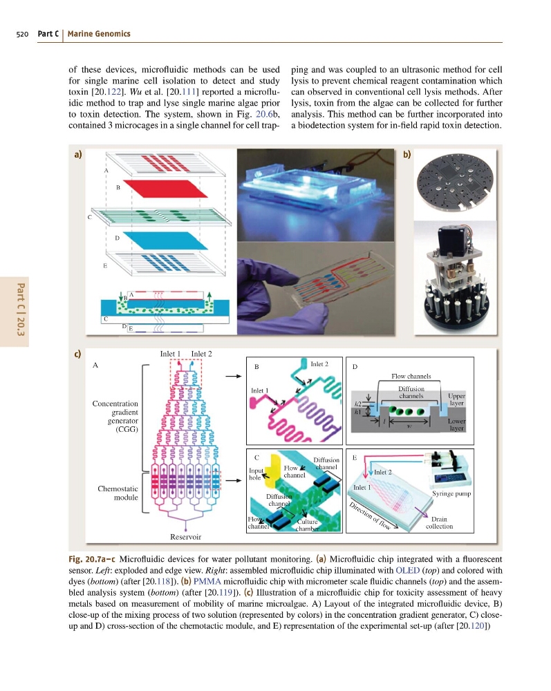
Fig. 20.7a–c Microfluidic devices for water pollutant monitoring. (a) Microfluidic chip integrated with a fluorescent
sensor. Left: exploded and edge view. Right: assembled microfluidic chip illuminated with OLED (top) and colored with
dyes (bottom) (after [20.118]). (b) PMMA microfluidic chip with micrometer scale fluidic channels (top) and the assembled analysis system (bottom) (after [20.119]). (c) Illustration of a microfluidic chip for toxicity assessment of heavy
metals based on measurement of mobility of marine microalgae. A) Layout of the integrated microfluidic device, B)
close-up of the mixing process of two solution (represented by colors) in the concentration gradient generator, C) closeup and D) cross-section of the chemotactic module, and E) representation of the experimental set-up (after [20.120])
ping and was coupled to an ultrasonic method for cell
lysis to prevent chemical reagent contamination which
can observed in conventional cell lysis methods. After
lysis, toxin from the algae can be collected for further
analysis. This method can be further incorporated into
a biodetection system for in-field rapid toxin detection.
520 Part C Marine Genomics
of these devices, microfluidic methods can be used
for single marine cell isolation to detect and study
toxin [20.122]. Wu et al. [20.111] reported a microfluidic method to trap and lyse single marine algae prior
to toxin detection. The system, shown in Fig. 20.6b,
contained 3 microcages in a single channel for cell trapa)
c)
b)
A
B
C
D
E
A
A
Concentration
gradient
generator
(CGG)
Chemostatic
module
Inlet 1
Inlet 1
Inlet 1
Inlet 2
Inlet 2
Inlet 2
B
D
C
E
Flow channels
Diffusion
channels
Upper
layer
Diffusion
channel
Diffusion
channel
Reservoir
Culture
chamber
Flow
channel
Drain
collection
D i r e c t i o n o f f l o w
Flow
channel
Input
hole
Lower
layer
Syringe pump
w
l
h2
h1
E
B
D
C
Fig. 20.7a–c Microfluidic devices for water pollutant monitoring. (a) Microfluidic chip integrated with a fluorescent
sensor. Left: exploded and edge view. Right: assembled microfluidic chip illuminated with OLED (top) and colored with
dyes (bottom) (after [20.118]). (b) PMMA microfluidic chip with micrometer scale fluidic channels (top) and the assembled analysis system (bottom) (after [20.119]). (c) Illustration of a microfluidic chip for toxicity assessment of heavy
metals based on measurement of mobility of marine microalgae. A) Layout of the integrated microfluidic device, B)
close-up of the mixing process of two solution (represented by colors) in the concentration gradient generator, C) closeup and D) cross-section of the chemotactic module, and E) representation of the experimental set-up (after [20.120])
ping and was coupled to an ultrasonic method for cell
lysis to prevent chemical reagent contamination which
can observed in conventional cell lysis methods. After
lysis, toxin from the algae can be collected for further
analysis. This method can be further incorporated into
a biodetection system for in-field rapid toxin detection.